


Physicians have separated a pair of conjoined twin girls who were born connected at the liver and, in a rare surgical case, at the heart, after completing a risky procedure made possible by 3-D printing.
In the operation, physicians at University of Florida Health Shands Children’s Hospital had to avoid damaging the critical veins in both babies’ hearts, as they were joined at the atrium— the upper chamber where the blood enters the organ— lead surgeon Dr. Mark Bleiweis said in a news release.
“In the world, there have not been many successful separations with a cardiac connection,” Bleiweis, chief of pediatric and congenital cardiovascular surgery at UF health, said in the release. “It became a very challenging planning process for us, and, ultimately, a challenging separation.”
(Courtesy University of Florida Health Shands Children’s Hospital)
According to UF Health, conjoined twins occur in about one in 200,000 live births, and survival of twins born connected at the heart is extremely rare. Often in these cases, doctors decide not to do separation surgery. Overall, only about 5 to 25 percent of conjoined twins survive.
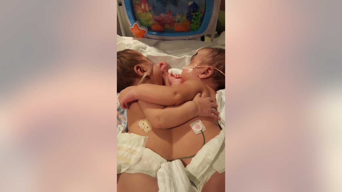
The twins at hand, named only as Scarlett and Savannah in the release, were born with a thoraco-omphalopagus connection, meaning they each had their own complete set of organs, but they were attached at not only the heart, but also the liver, the diaphragm and the sternum.
Dr. Jennifer Co-Vu, director of the fetal cardiac program at Shands, began studying the twins’ physiology while their mother, named only as Jacquelyn in the release, was 21 weeks pregnant. Jacquelyn said when she learned her twins were conjoined at 20 weeks pregnant, her previous doctor told her the babies likely would not survive.
“We went in to find the sex of one baby and found out not only were they twins, but they were conjoined and weren’t going to make it,” Jacquelyn said in the release. “Many opinions later, we found Dr. Co-Vu, and she told us not only were our twins going to live, but they thought they could separate them.”
Co-Vu and her team produced what they said appeared to be the first 3-D printed conjoined twin heart to help the surgical team plan and practice for the operation.
“When I saw the heart structures and liver structures in utero, I had a feeling that we could separate them, but I had to examine the anatomy more closely and consult with my cardiology colleagues at the UF Health Congenital Heart Center,” Co-Vu said in the release. “I was able to give them hope, yet at the same time, I told them I was cautiously optimistic. … We are very fortunate that this was a success.”
Dr. Saleem Islam, chief of the division of pediatric surgery in the UF College of Medicine, used an intraoperative ultrasound to navigate separation of the newborns’ single large, fused liver. The technology helped Islam and his team identify parts of the liver where large blood vessels were absent.
In a six- to eight-hour surgery, four surgeons and two anesthesiologists, as well as a team of imaging specialists, multiple nurses and an ancillary staff successfully separated the infants. According to the release, physicians wrapped the twins with tubing and electrical wiring, and to be able to tell them apart, they wrapped orange tape on one of the twins and blue tape on the other.
After their babies have undergone more than a dozen surgeries each, Jacquelyn and her partner Mark are preparing to bring them home.
“I know that Mark and Jackie were told by many not to pursue this because it was daunting, and it could not and would not be successful,” Bleiweis said in the release. “Nothing gives us greater satisfaction than seeing the two twins separated, and to see both parents holding their twins.”