


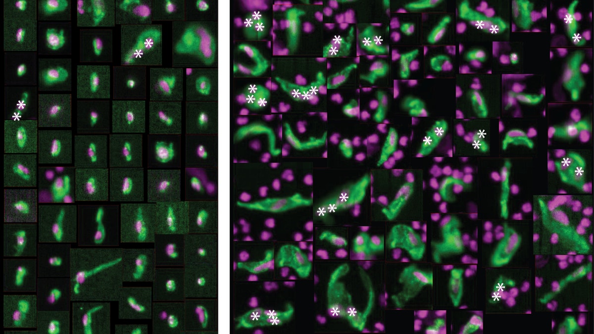
On the left are normal, healthy cells that line the coronary artery -- also known as circulating endothelial cells (CEC). On the right are CECs from heart attack patients which appear abnormally large, misshapen and with multiple nuclei.
It can happen so fast.
First, a person experiences chest pain, and suddenly he or she is in the emergency room – fighting for life after a massive heart attack. In some cases, it seems to come out of nowhere.
But what if there were a way to predict whether or not you will have a heart attack? Now, a foretelling test could soon be a reality.
A study from the Scripps Translational Science Institute (STSI) in La Jolla, Calif., has revealed a potential blood test that ER doctors can use to determine if patients visiting the hospital complaining of chest pains may soon be back due to suffering a life-threatening heart attack. The blood test involves examining specific cells from a person’s coronary artery, which were found to be abnormally large and misshapen in the days before an impending heart attack.
“This is a critical unmet need in medicine,” Dr. Eric Topol, the study’s lead investigator and director of STSI, told FoxNews.com. “To be able to know before a patient has a heart attack to prevent a clot from forming. We have potent medicines to [treat clots], but we don’t know which people to treat.”
Currently, there are a number of tests doctors perform on patients who check into the ER for chest pains – including a troponin test, a CK-MB, or a cardiogram. Unfortunately, these tests only look for pre-existing damage to a person’s arteries and can only determine if a patient is currently experiencing a heart attack or have recently experienced a heart attack. But many patients who go to the hospital with chest discomfort show no signs of artery damage and are sent home - only to have a heart attack within the next few days.
“Their [coronary] artery has been under intense inflammation, and that happens for days before a blood clot forms to seal up the crack that forms [in the artery],” Topol said. “And that’s the heart attack, when there’s a cut off of the artery where the clot forms.”
In order to reveal inflammation in a person’s artery, Topol and his team studied 50 patients in the early minutes of a heart attack. They took samples of their blood and isolated the cells that were coming from inside their artery – cells known as circulating endothelial cells (CEC).
Typically these cells act like Saran Wrap – they are tightly banded together and wrapped around the artery in order to protect it. But as the artery becomes inflamed, the CECs begin to shed and start to circulate throughout the bloodstream – a place they aren’t supposed to be. And according to this latest study, they don’t look the way that they should.
“Not only were there 400 percent more CECs in these people [suffering heart attacks], but also the cells were very unique,” Topol said. “Typically a cell like this is elliptical in shape. But these were in big clusters; giant cells with multiple nuclei. A very different signature compared to what’s normal.”
Topol compared the CECs to a control group of 44 healthy people, all of which had normal CEC counts and shapes. Because of the comparison, the team concluded that CECs are reliable biomarker that can safely determine a developing heart attack.
According to Dr. Paddy Barrett, a lead investigator at STSI, this discovery could potentially help over 2.5 million Americans who experience a heart attack brought on by obstructive coronary artery disease – when a clot forms to plug a cracked artery. With the possibility to save so many lives on the horizon, the team is already in the process of developing an inexpensive test to be used by emergency room doctors.
And they expect to have it available in the next one to two years.
“We can finally be more precise,” Topol said. “Now there’s the heart attack department and the pre-heart attack department. So it’s a whole different ability to diagnose. It’s digitizing the cells in the blood to diagnose a heart attack in the making.”