


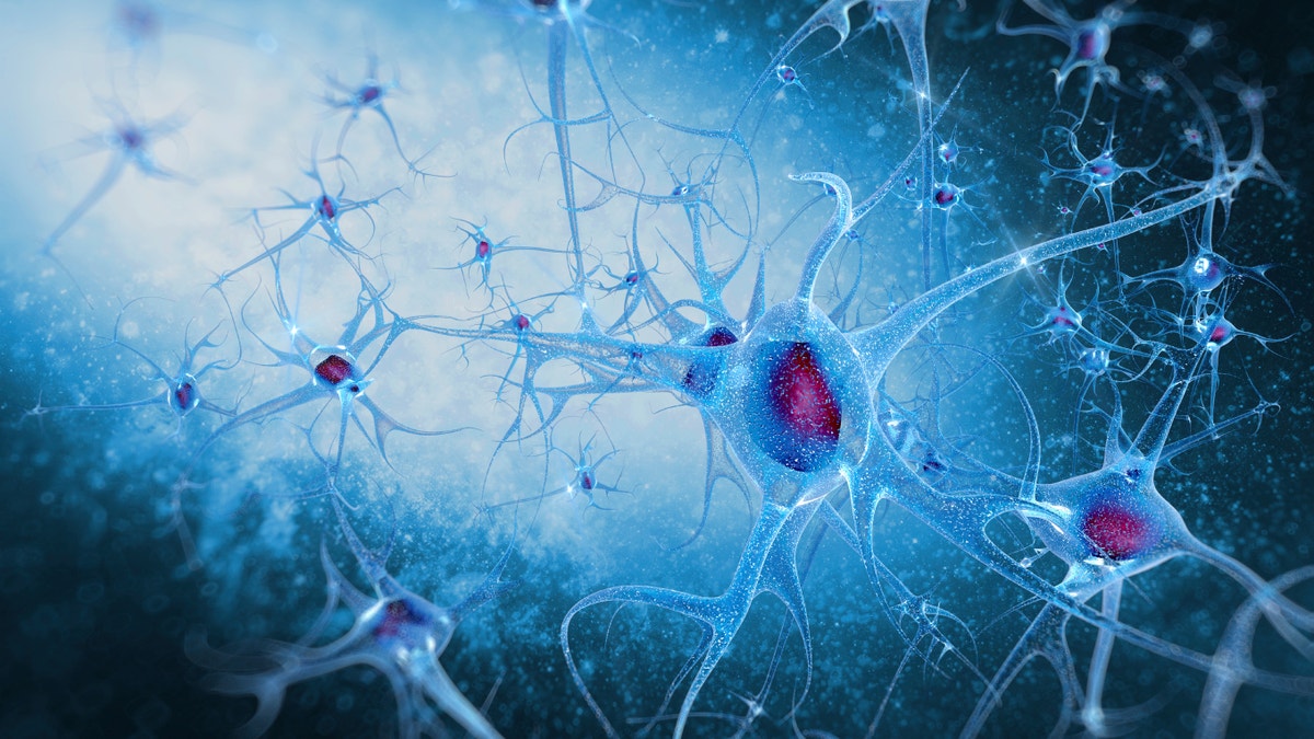
WASHINGTON — The brain's nerve cells communicate by firing messages to each other through junctions called synapses, and problems with those connections are linked to disorders like Alzheimer's and epilepsy. Now Yale University researchers have developed a way to picture synapses in living brains.
The technique reported Wednesday, using PET scans, is highly experimental but it raises the possibility of one day monitoring synapse function in some common diseases.
A healthy human brain harbors trillions of synapses, a number that changes over a lifetime.
Early in life, the brain "prunes" the many synapses between neurons so the right number is in each region, a process that can go wrong in disorders such as autism or schizophrenia. Changes in the density of synapses may signal where epilepsy seizures originate. Later in life, synapse loss is associated with Alzheimer's disease.
But measuring synapses has required autopsies, or occasional attempts during brain surgery.
To find a non-invasive approach, the Yale-led team developed a radioactive compound, called a tracer, that is injected into the body and binds with a particular protein that is found in the brain's synapses. The idea: During a PET scan, those synapses appear lit up against dark, synapse-free areas of the brain.
Animal testing confirmed the tracer was targeting synapses.
The research team then mapped the density of synapses in the brains of 10 healthy volunteers and three patients with a form of epilepsy. Compared to the healthy brains, the technique revealed lost synapses in the epilepsy-affected regions of those patients' brains, the researchers reported Wednesday in the journal Science Translational Medicine.
"This work represents a breakthrough in the ability to study an important process in the brain that is not only part of normal brain development but that also may be involved in several neuropsychiatric diseases," said Dr. Peter Herscovitch, who directs PET scanning at the National Institutes of Health's Clinical Center and wasn't involved in the research.
Much more work is needed to make the tracer last longer in the brain, a key if it's ever to be of use to doctors, cautioned Yale radiology professor Richard Carson, the study's senior author. But even though it starts disappearing quickly, he said it's a good tool to research brain function.
Stay tuned: Carson's team has begun using the technique to study Alzheimer's, to determine if changes in synaptic density over time can help predict that disease's development.