


Skin biopsy without cutting
Dermatologists usually do a biopsy if you have a suspicious spot on your body. Now new technology gets results quicker, without cutting
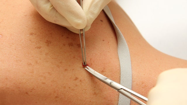
If you have a suspicious spot on your body, dermatologists usually do a biopsy to determine if it is cancer. That means numbing, cutting and waiting for your results for up to two weeks.
But now, doctors are using new technology that is non-invasive and much quicker: confocal microscopy.
“It is a device like a camera, which takes images of skin lesions, which is then displayed on a computer screen, where you can see actual cellular details to see if the condition is benign or not benign,” said Dr. Babar Rao, director of Rao Dermatology in New York City.
Confocal microscopy has been around for more than 10 years, but it’s only been used for research – until now. According to Rao, the entire process is very quick.
"We can scan the whole lesion within several minutes,” Rao said. “We can go side to side and take as many pictures as you like."
The images of the skin lesions are then magnified so that doctors can look for any abnormal cells.
Fortunately, for 3-year-old Katie Strong, the process showed her lesion to be benign.
“It's easy; it's painless,” said Jennifer Strong, Katie’s mother. “…It took less than a minute, and now we know that (the lesion is) okay, and we don't have to take it off."
Confocal microscopy costs about $250 dollars and is not covered by insurance. Rao said this procedure is ideal if the patient has a mole on his or her face or breast.