


(Flickr/illuminaut)
What if teachers could predict a child’s special needs and develop a unique educational program in advance? And what if doctors could know whether a psychiatric medication would worsen or improve a particular patient’s symptoms?
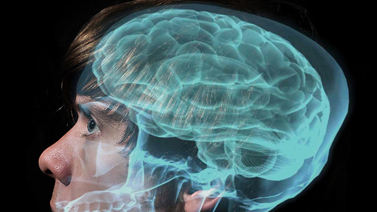
Researchers at the Massachusetts Institute of Technology (MIT) have compiled dozens of studies that suggest analyzing data from brain imaging, such as functional magnetic resonance imaging (fMRI), may one day help scientists make these predictions— and more. Their review, which features medical research from the past 14 years, includes findings that link specific neuromarkers to a person’s likelihood of participating in criminal activity, an individual’s potential success with a drug or alcohol treatment, and someone’s likelihood of developing a certain talent over his or her lifetime.
“Seventy or so studies have reported positive findings that analyzing brain measures beforehand can considerably improve knowing whether a person will be successful at something,” lead study author John Gabrieli, a neuroscience professor at MIT, told FoxNews.com. “In many areas in medicine or education it’s purely a guessing game.”
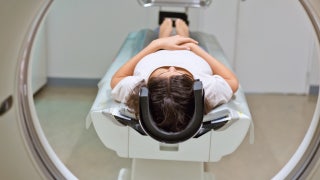
Brain imaging is a noninvasive method for measuring brain activity. For example, fMRI— the primary brain imaging method used in the review studies— measures brain activity associated with changes in blood flow that occur in response to neural activity. The review also includes studies that use electroencephalography (EEG), a measurement of electrical activity in the brain that is gathered by recording from electrodes placed on the scalp.
Study author Susan Whitfield-Gabrieli, a brain research scientist at MIT, said only half of the treatments for mental disorders— such as anxiety and depression— prove effective, and many doctors misdiagnose based on a lack of individual patient information. She noted that there is a wide variety of depression medication, and an inappropriate prescription can result in uncomfortable side effects, frustrated attrition or, worst-case scenario, fatality.
“Right now, physicians don’t have any objective mechanism to determine which therapy will benefit which patient,” Whitfield-Gabrieli said. “Our hope is to use brain imaging to help with better diagnosis.”
Most behavioral issues are analyzed based on patient observation and self-reporting through surveys and quizzes, but these methods can fall short. Brain imaging often outperforms those assessments or can serve to buttress them, the current review illustrates.
In a 2011 study of children with dyslexia, behavioral measures didn’t correlate with an individual’s reading gains over two and a half years, but brain measures did. Two different neuromarkers predicted with 72 percent accuracy whether a child would see his or her reading skills stagnate or decline over the study period.
Research in 2007 similarly focused on trends among children identified as struggling readers over a one-year period. Beginning-of-year behavioral measures accounted for 65 percent of the variance in end-of-year reading scores, and brain measures accounted for only 57 percent of that variation. But taken together, the behavioral and brain measures accounted for 81 percent of the variance, indicating the strongest forecast of student reading skills over a school year.
Providing the most accurate diagnosis for autism spectrum disorder (ASD) requires brain imaging techniques, as many children on the spectrum may not be able to perform a given behavioral task, Whitfield-Gabrieli pointed out. With resting state connectivity, a technology that doesn’t require the subject to complete a task, researchers can examine detailed brain maps of children who may have ASD.
Study authors noted that one limitation of studies conducted to date, including the research referenced in their paper, is a modest study population size. The largest study included in the review includes about 270 participants, but most of the featured studies consist of fewer than 100.
To effectively use brain imaging as a means for predicting future behavior— and eventually supply the best intervention recommendation— larger studies must first be conducted, they noted.
The single study in the review that focuses on criminal behavior, for example, followed 96 male offenders over a four-year period. The research, published in 2013, suggested that the likelihood of an offender being rearrested over the study period doubled when, at baseline, the person had low activation in the anterior cingulate cortex— a region linked to cognitive control and specifically the resolution of cognitive conflict.
The research, like much of the relevant studies conducted to date, suggests another limitation: a simple correlation between neuromarkers and behavior. Brain imaging is not predictive by definition because the knowledge of these analyses hinges on their outcomes, the study authors write in their paper, which was published Wednesday in the journal Neuron.
In addition to examining larger study groups, using brain imaging data to predict behavior will also require using a heterogeneous rather than a homogenous model, study author Satrajit Ghosh, a brain imaging research scientist at MIT, told FoxNews.com. The current study relies on the homogenous model, which analyzes brain differences within a single study group.
“We want to be able to say something with confidence about an individual person,” Ghosh said, “and many times when we look at these studies, we look at relations within a group, and we say, ‘It looks like this treatment was effective and why.’ But what we want to do in terms of personalizing medicine is to say, ‘How well will it do on any given individual?’ So we need to be able to generalize an ‘out of sample’ prediction— or create a model based on a set of people and then use that model to create a plan for someone not involved in a study.”
Using brain imaging data to make predictions and help solve problems before they arise is the ultimate goal, researchers noted.
“It’s harder to rescue somebody than it is to give them support,” Gabrieli said.