


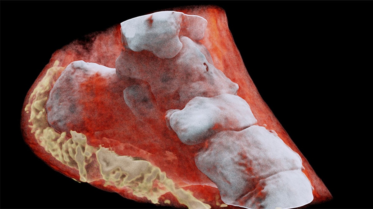
A magnified X-ray captured by CERN's Medipix. (mars bioimaging.com)
Scientists from New Zealand recently performed the first-ever 3-D, color X-ray on a human being.
Developed by the European Organization for Nuclear Research, known as CERN, the new device works like a camera, collecting individual sub-atomic particles as they rapidly collide with pixels when its shutter is open, the Agence France Press reported.
This allows for high-contrast, high-resolution pictures to be produced at a quick rate.
The device, named “Medipix,” incorporates particle-tracking technology developed for CERN's Large Hadron Collider, which in 2012 discovered the Higgs Boson particle.
“This color X-ray imaging technique could produce clearer and more accurate pictures and help doctors give their patients more accurate diagnoses,” read a statement from CERN.
The “Medipix” is unique because it has the capability to show practitioners the difference between bone, muscle and cartilage in real time.
With this degree of advanced technology, doctors can examine the exact positioning and size of cancerous tumors, which will help to improve the quality of physical operation as we know it.
This technology is now being commercialized by New Zealand company MARS Bioimaging Ltd, which is linked to two universities that helped develop it: Otago and Canterbury.