Move Back
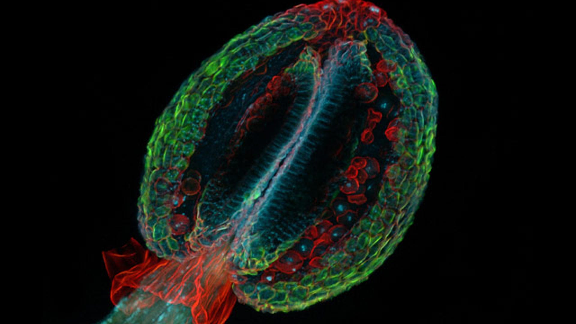
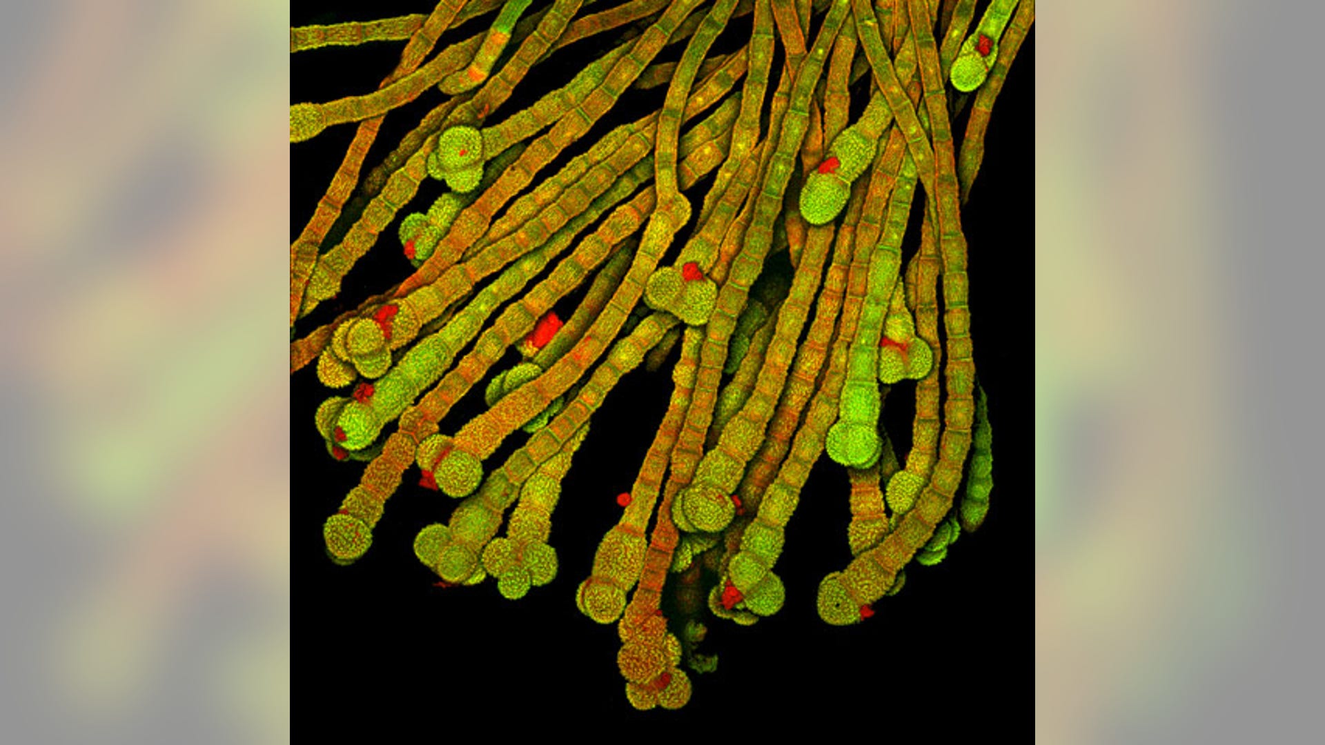
![1. Thale Cress]() Arabidopsis thaliana is the first plant to have its genome fully sequenced and is commonly used as a model in scientific research. But it was the unusually artistic appearance of the winning shot that inspired photomicrographer and plant biologist Dr. Heiti Paves of the Tallinn University of Technology in Estonia to enter the image into the 35-year-old competition. According to Dr. Paves, besides being "nice-looking plant organs," anthers were a good subject because "they do not move very fast… The picture of my dreams should bring out motility of living cell, like a sports photograph."
Arabidopsis thaliana is the first plant to have its genome fully sequenced and is commonly used as a model in scientific research. But it was the unusually artistic appearance of the winning shot that inspired photomicrographer and plant biologist Dr. Heiti Paves of the Tallinn University of Technology in Estonia to enter the image into the 35-year-old competition. According to Dr. Paves, besides being "nice-looking plant organs," anthers were a good subject because "they do not move very fast… The picture of my dreams should bring out motility of living cell, like a sports photograph."![2. Flower Stem]() Not all of the winning images were created by scientists using expensive state-of-the-art equipment. Gerd A. Guenther is an organic farmer from Dusseldorf, Germany where he produces vegetables, potatoes and hay for horses. His stunning picture of a thin cross section of the stem of a Sonchus asper blossom, a yellow blooming wildflower often found on farmland, won second prize. The plant was magnified 150 times, bringing a new perspective to the wonders of nature. "The remarkable contrast between the red hats of the plant hairs and the green stem in combination with the white stems thrilled me," said Mr. Guenther.
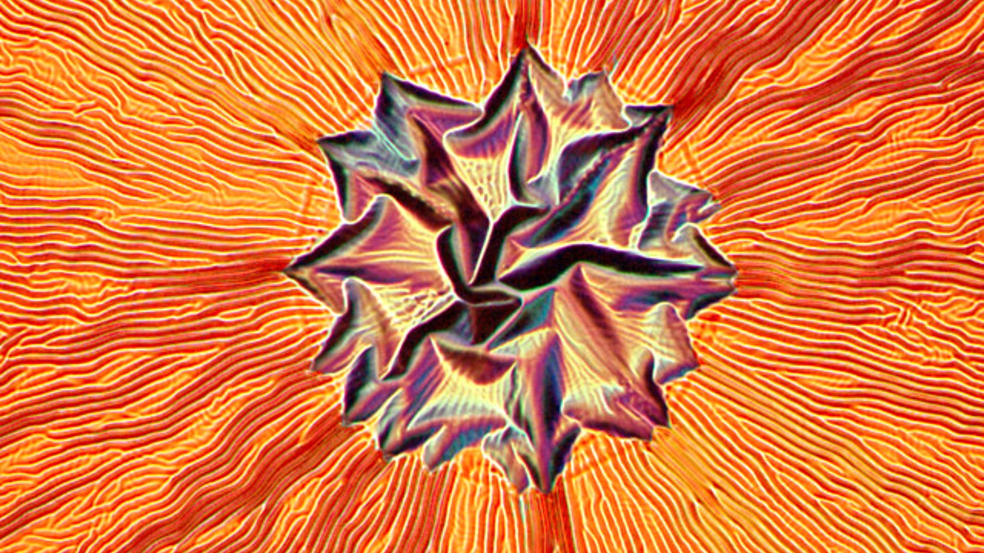
Not all of the winning images were created by scientists using expensive state-of-the-art equipment. Gerd A. Guenther is an organic farmer from Dusseldorf, Germany where he produces vegetables, potatoes and hay for horses. His stunning picture of a thin cross section of the stem of a Sonchus asper blossom, a yellow blooming wildflower often found on farmland, won second prize. The plant was magnified 150 times, bringing a new perspective to the wonders of nature. "The remarkable contrast between the red hats of the plant hairs and the green stem in combination with the white stems thrilled me," said Mr. Guenther.![3. Wrinkled Seimiconductor]() Dr. Pedro Barrios-Perez used brightfield to capture the wrinkled photoresist magnified 200 times in his winning image. Dr. Barrios-Perez of the Institute for Microstructural Sciences at the National Research Council of Canada in Ottawa, won third place with a failed attempt to develop a photoresist pattern on a semiconductor. "These pictures are taken out of my interest in art," said Dr. Barrios-Perez. "If I show it to my boss, he just says, 'Throw the sample away.' I thought that it looked like a face with a fire that was warming up my days." He added that the particular result "cannot be reproduced – some of this stuff just happens."
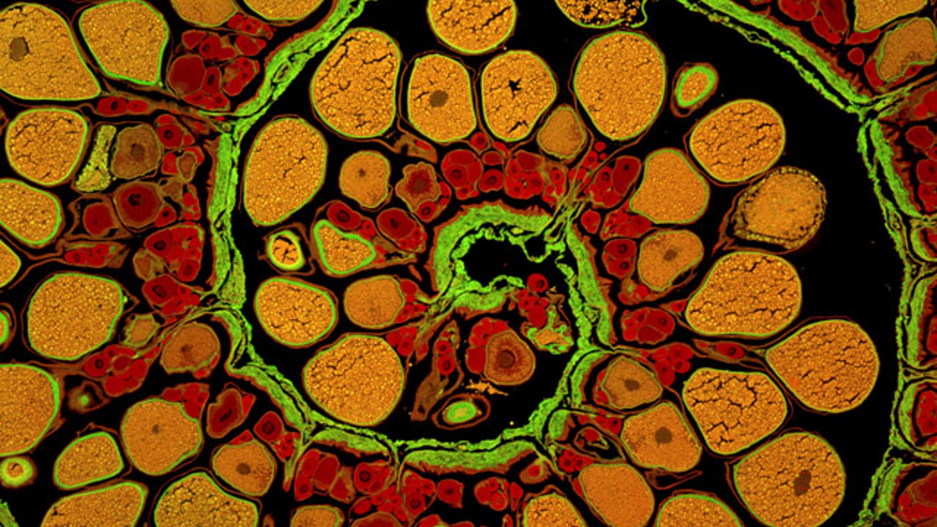
Dr. Pedro Barrios-Perez used brightfield to capture the wrinkled photoresist magnified 200 times in his winning image. Dr. Barrios-Perez of the Institute for Microstructural Sciences at the National Research Council of Canada in Ottawa, won third place with a failed attempt to develop a photoresist pattern on a semiconductor. "These pictures are taken out of my interest in art," said Dr. Barrios-Perez. "If I show it to my boss, he just says, 'Throw the sample away.' I thought that it looked like a face with a fire that was warming up my days." He added that the particular result "cannot be reproduced – some of this stuff just happens."![4. Anglerfish ovary]() When a former colleague sent him a section of an anglerfish ovary, James E. Hayden of The Wistar Institute came up with the idea of looking at the autofluorescence of the tissue in two colors. His vibrant swirling photomicrograph of developing oocytes, or unfertilized eggs, as they move along the spiral of an anglerfish's ovary came in fourth. Mr. Hayden said he is drawn to both photographic art and science. "Most microscopists have a streak of artist in them. It's hard not to. You're looking at things through a microscope that most people don't see. The nascent artist in you sort of peeks its head up."
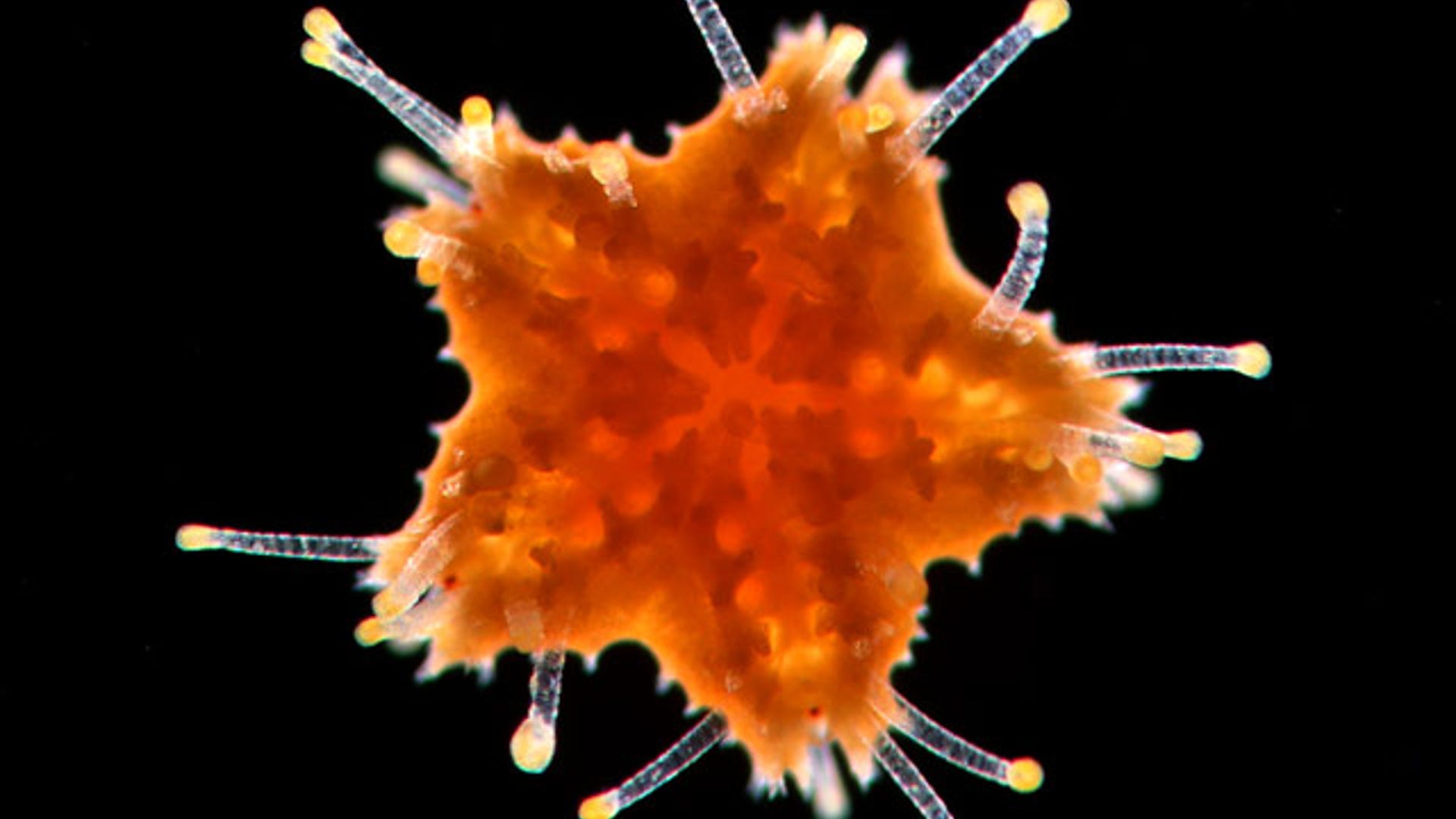
When a former colleague sent him a section of an anglerfish ovary, James E. Hayden of The Wistar Institute came up with the idea of looking at the autofluorescence of the tissue in two colors. His vibrant swirling photomicrograph of developing oocytes, or unfertilized eggs, as they move along the spiral of an anglerfish's ovary came in fourth. Mr. Hayden said he is drawn to both photographic art and science. "Most microscopists have a streak of artist in them. It's hard not to. You're looking at things through a microscope that most people don't see. The nascent artist in you sort of peeks its head up."![5. Young Seastar]() A young, hungry sea star appears to open its mouth wide as its transparent tube feet grasp for morsels. Marine biologist Bruno Vellutini of the University of Sao Paulo in Brazil said the sea star, or starfish, was imaged at 40 times magnification shortly after it had metamorphosed into a juvenile. He said he was lucky to find the juvenile seastar in the plankton samples he collected while looking for sand dollar larvae for his master’s thesis last year. "Scientific images don’t need to be beautiful," he said. "To take a good picture, you need patience to prepare light, make sure everything is clean. It takes a lot of effort to create a technically nice image." But, he said, taking beautiful pictures "makes research more fun."
A young, hungry sea star appears to open its mouth wide as its transparent tube feet grasp for morsels. Marine biologist Bruno Vellutini of the University of Sao Paulo in Brazil said the sea star, or starfish, was imaged at 40 times magnification shortly after it had metamorphosed into a juvenile. He said he was lucky to find the juvenile seastar in the plankton samples he collected while looking for sand dollar larvae for his master’s thesis last year. "Scientific images don’t need to be beautiful," he said. "To take a good picture, you need patience to prepare light, make sure everything is clean. It takes a lot of effort to create a technically nice image." But, he said, taking beautiful pictures "makes research more fun."![6. Discus Fish Scales]() Discus fish scales, magnified 20X, as captured by Havi Sarfaty of the Israel Veterinary Association.
Discus fish scales, magnified 20X, as captured by Havi Sarfaty of the Israel Veterinary Association.![7. Black-Eyed Susan Vine]() The hair-like trichomes on Thunbergia alata, more commonly called the Black-eyedSusan vine, magnified 450X.
The hair-like trichomes on Thunbergia alata, more commonly called the Black-eyedSusan vine, magnified 450X.![8. Cotton Fibers]() Cotton fibers stained with berberine sulphate and shaded to indicate depth, magnified 200X, explain this stunning image.
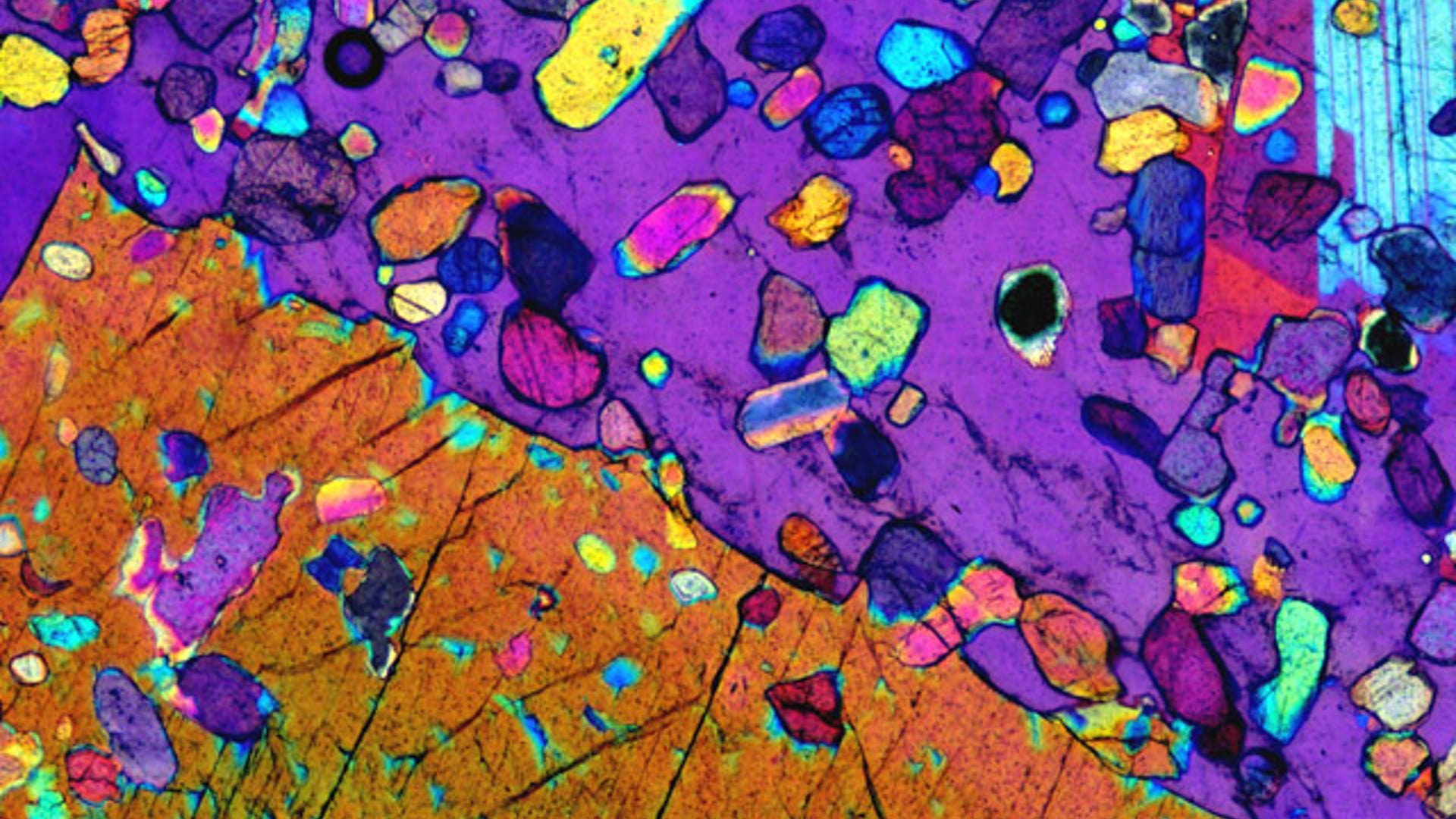
Cotton fibers stained with berberine sulphate and shaded to indicate depth, magnified 200X, explain this stunning image.![9. Magmatic Rock]() Inclusions are materials trapped within minerals during their formation.Here, Olivine inclusions in gabbro (magmatic rock), after being magnified 5X.
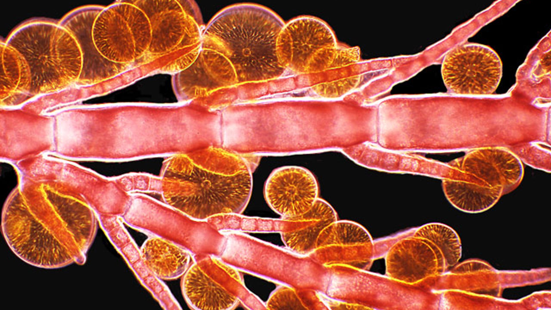
Inclusions are materials trapped within minerals during their formation.Here, Olivine inclusions in gabbro (magmatic rock), after being magnified 5X.![10. Algae and diatoms]() Algae and diatoms at 10X magnification.>
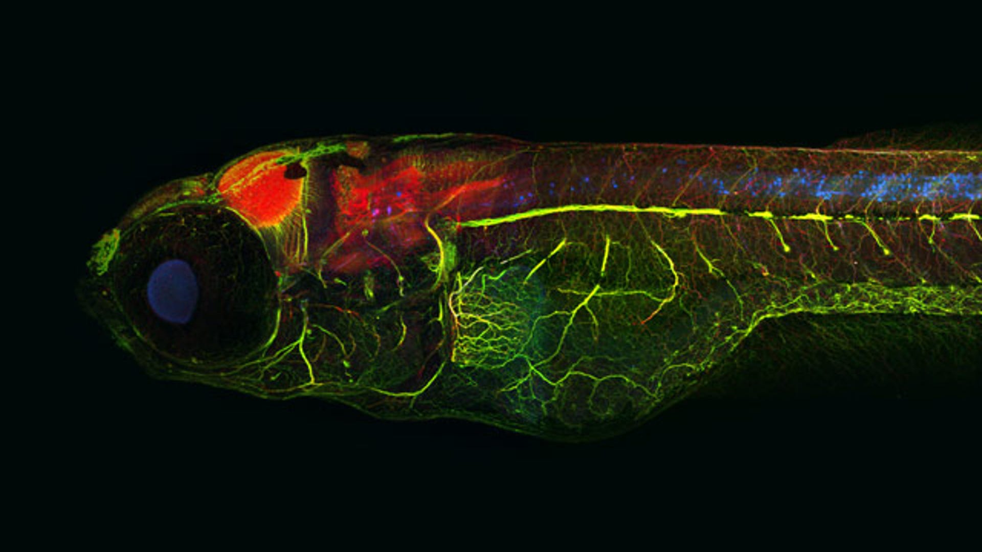
Algae and diatoms at 10X magnification.>![11.]() "Alzheimer" Zebrafish, stained for Tau (red), neurons (green), and pathologic Tau (blue), at a 10X magnification.
"Alzheimer" Zebrafish, stained for Tau (red), neurons (green), and pathologic Tau (blue), at a 10X magnification.![12. Soap Film]() Tsutomu Seimiya captured this picture of the flow pattern in draining soap film at 10X magnification.
Tsutomu Seimiya captured this picture of the flow pattern in draining soap film at 10X magnification.![13. Recrystalized Chemicals]() A recrystallized melted mixture of acetanalide, resorcinal and carbon tetrabromide chemical,s magnified 33X.
A recrystallized melted mixture of acetanalide, resorcinal and carbon tetrabromide chemical,s magnified 33X.![14. Lobster Egg]() Tora Bardal of Norway's NTNU Center of Fisheries and Aquaculture captured this photo of a lobster egg, magnified 3.2X.
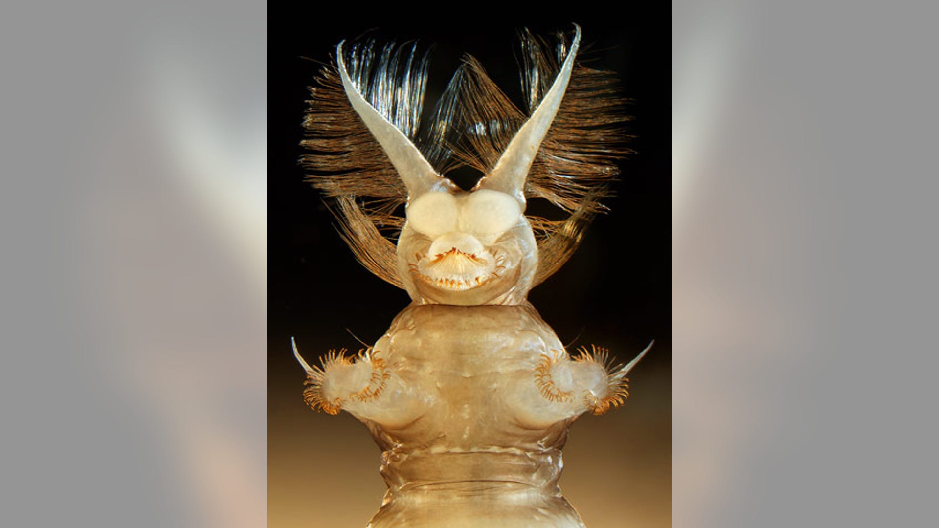
Tora Bardal of Norway's NTNU Center of Fisheries and Aquaculture captured this photo of a lobster egg, magnified 3.2X.![15. Fly Larva]() What is it? Why Atherix ibis, the aquatic larva of a a fly magnified 25X.
What is it? Why Atherix ibis, the aquatic larva of a a fly magnified 25X.![16. Snail Eggs]() Snail eggs (Lymnaea sp.) at 200X, as photographed by Massimo Brizzi of Microcosmo Italia.
Snail eggs (Lymnaea sp.) at 200X, as photographed by Massimo Brizzi of Microcosmo Italia.![17. Stopwatch]() An ordinary stopwatch takes on vivid hues thanks to depth coding and a 2.5x magnification.
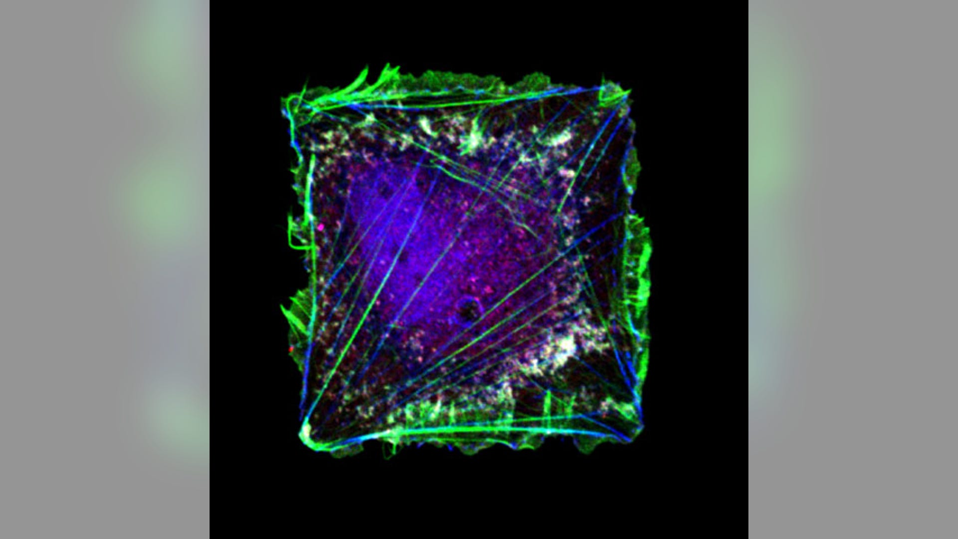
An ordinary stopwatch takes on vivid hues thanks to depth coding and a 2.5x magnification.![18. Human Skin]() Human skin on fibronectin with growth factor, shown magnified at6 60X.
Human skin on fibronectin with growth factor, shown magnified at6 60X.![19. Snowflake]() Not your ordinary snowflake, as photographed by Yanping Wang of the Beijing Planetarium.
Not your ordinary snowflake, as photographed by Yanping Wang of the Beijing Planetarium.![20. Rusted Old Coin]() A rusted old coin, photographed at 40x magnification.
A rusted old coin, photographed at 40x magnification.Beauty Beneath the Microscope's Lens
Nikon's Small World contest showcases the beauty of life as photographed through a microscope. Here, the winners from 2009's contest.


Move Forward
- Beauty Beneath the Microscope's Lens


