Move Back
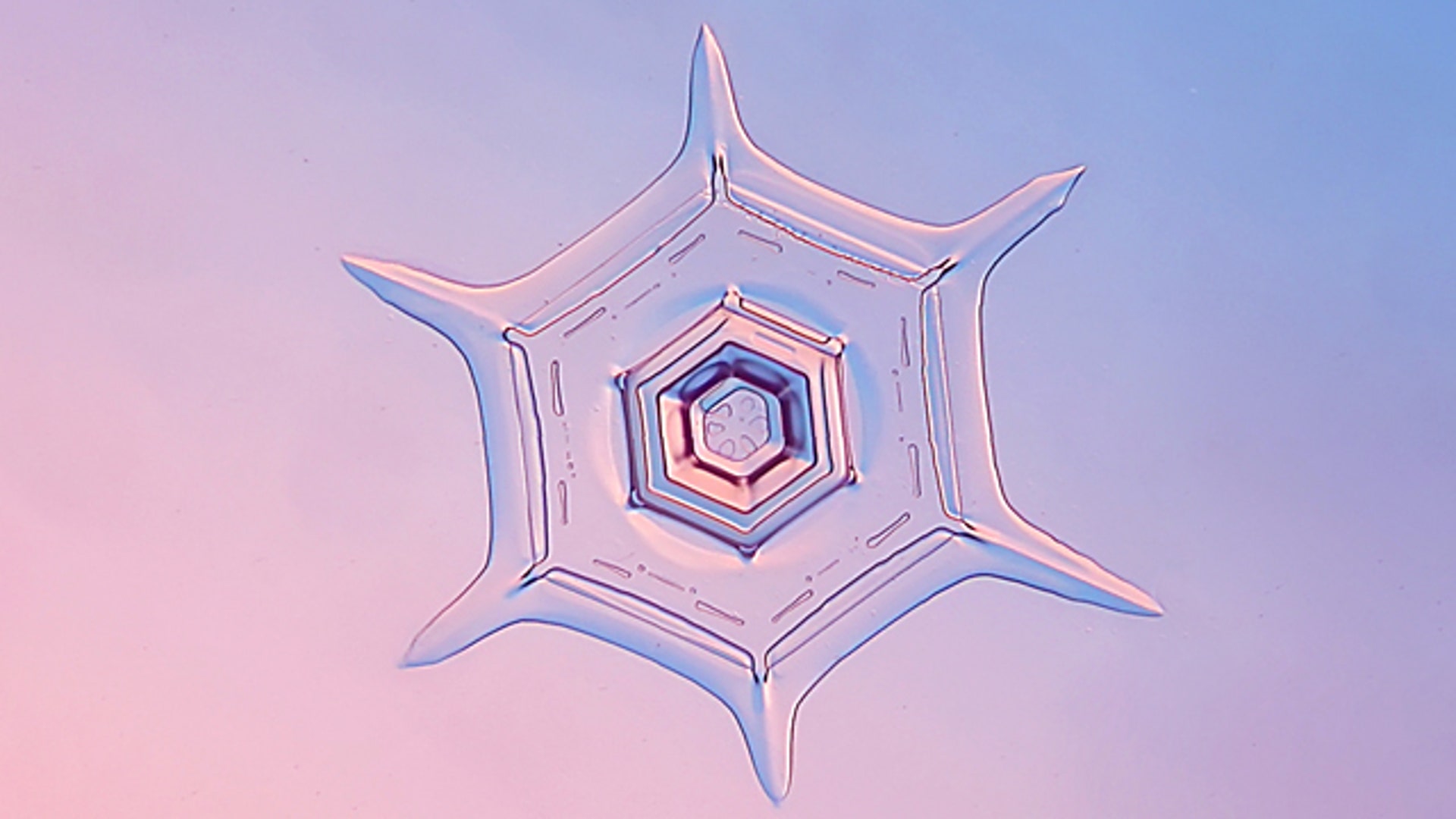
![Snowflake]() A photomicrograph is a technical document that can be of great significance to science. A one is also an object of beauty, open to several levels of appreciation. And once a year, Nikon celebrates the best of the bunch. Pictured here: a snowflake magnified 4X by Yangping Wang of the Beijing Planetarium. It received an honorable mention.
A photomicrograph is a technical document that can be of great significance to science. A one is also an object of beauty, open to several levels of appreciation. And once a year, Nikon celebrates the best of the bunch. Pictured here: a snowflake magnified 4X by Yangping Wang of the Beijing Planetarium. It received an honorable mention.![1 Igor Siwanowicz]() 1st Place -- Dr. Igor Siwanowicz Portrait of a green lacewing larva Technique: Confocal, magnified 20X
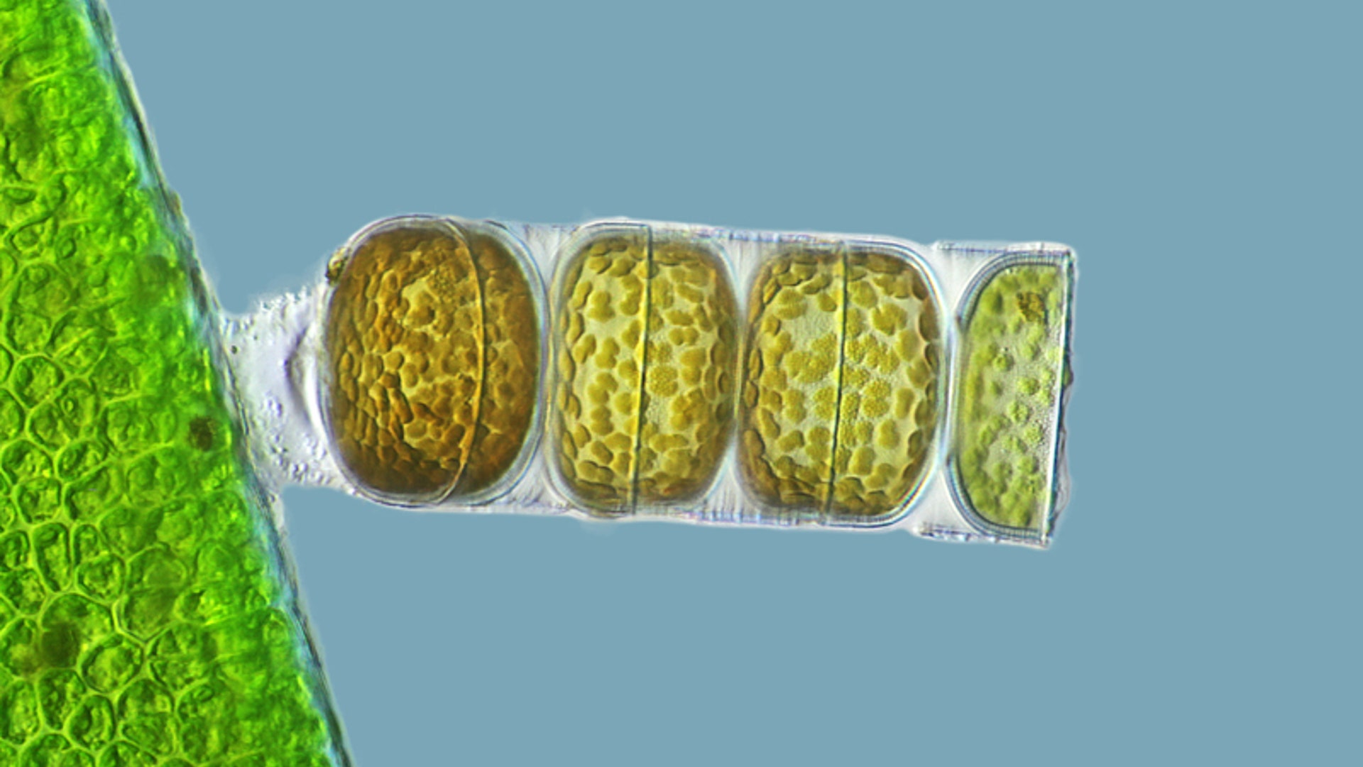
1st Place -- Dr. Igor Siwanowicz Portrait of a green lacewing larva Technique: Confocal, magnified 20X![2 Donna Stolz]() 2nd Place -- Dr. Donna Stolz Blade of grass Technique: Confocal stack reconstruction, autofluorescence, magnified 200X
2nd Place -- Dr. Donna Stolz Blade of grass Technique: Confocal stack reconstruction, autofluorescence, magnified 200X![3 Frank Fox]() 3rd Place -- Frank Fox Melosira monoliformis (living specimen) Technique: Differential interference contrast, magnified 320X
3rd Place -- Frank Fox Melosira monoliformis (living specimen) Technique: Differential interference contrast, magnified 320X![4 Robin Young]() 4th Place -- Dr. Robin Young Liverwort Technique: Live mount, confocal microscopy, magnified 20X
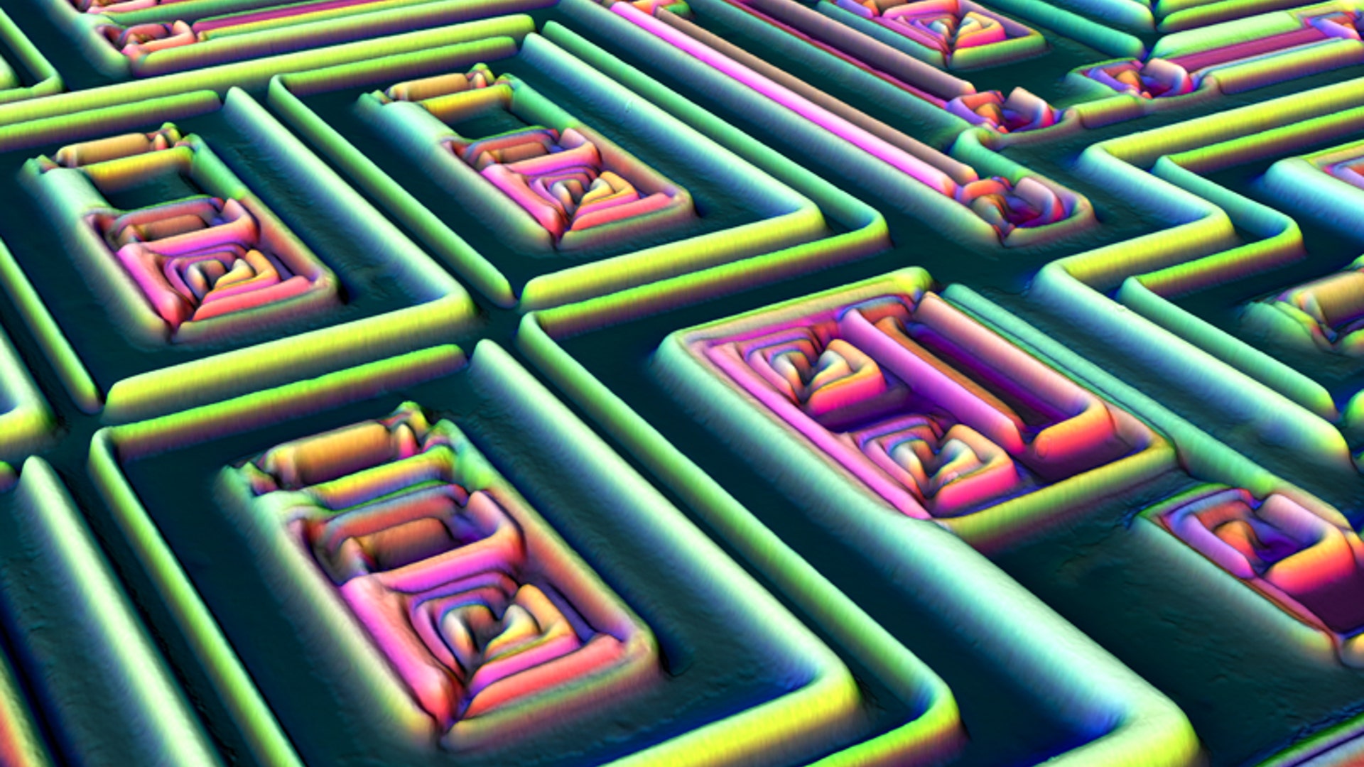
4th Place -- Dr. Robin Young Liverwort Technique: Live mount, confocal microscopy, magnified 20X![5 Alfred Pasieka]() 5th Place -- Alfred Pasieka 3D reconstruction of a microchip Technique: Incident light, Normarski interference contrast, magnified 500X
5th Place -- Alfred Pasieka 3D reconstruction of a microchip Technique: Incident light, Normarski interference contrast, magnified 500X![6 Dennis Callahan]() 6th Place -- Dennis Callahan Cracked solar cell films Technique: Brightfield, magnified 50X
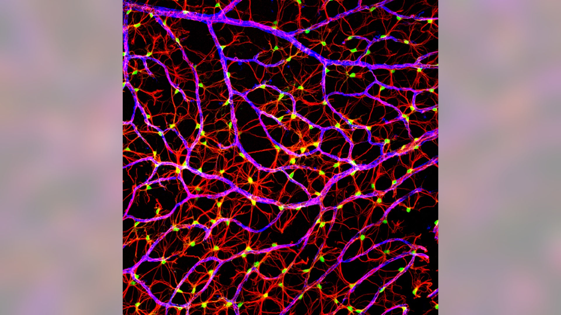
6th Place -- Dennis Callahan Cracked solar cell films Technique: Brightfield, magnified 50X![7 Gabriel Luna]() 7th Place -- Gabriel Luna Nerve fibers from the retina of a mouse Technique: Laser confocal scanning, magnified 40X
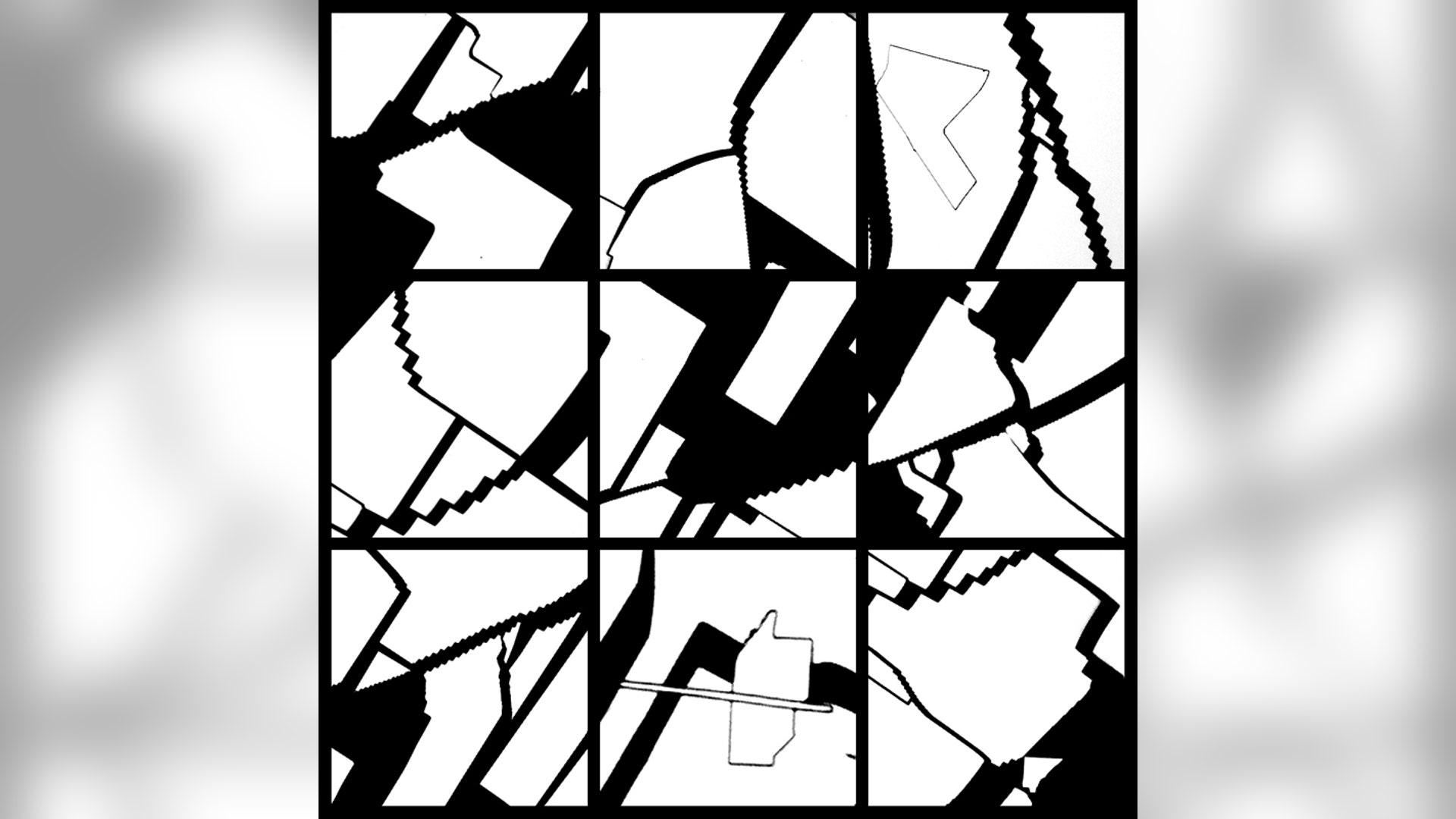
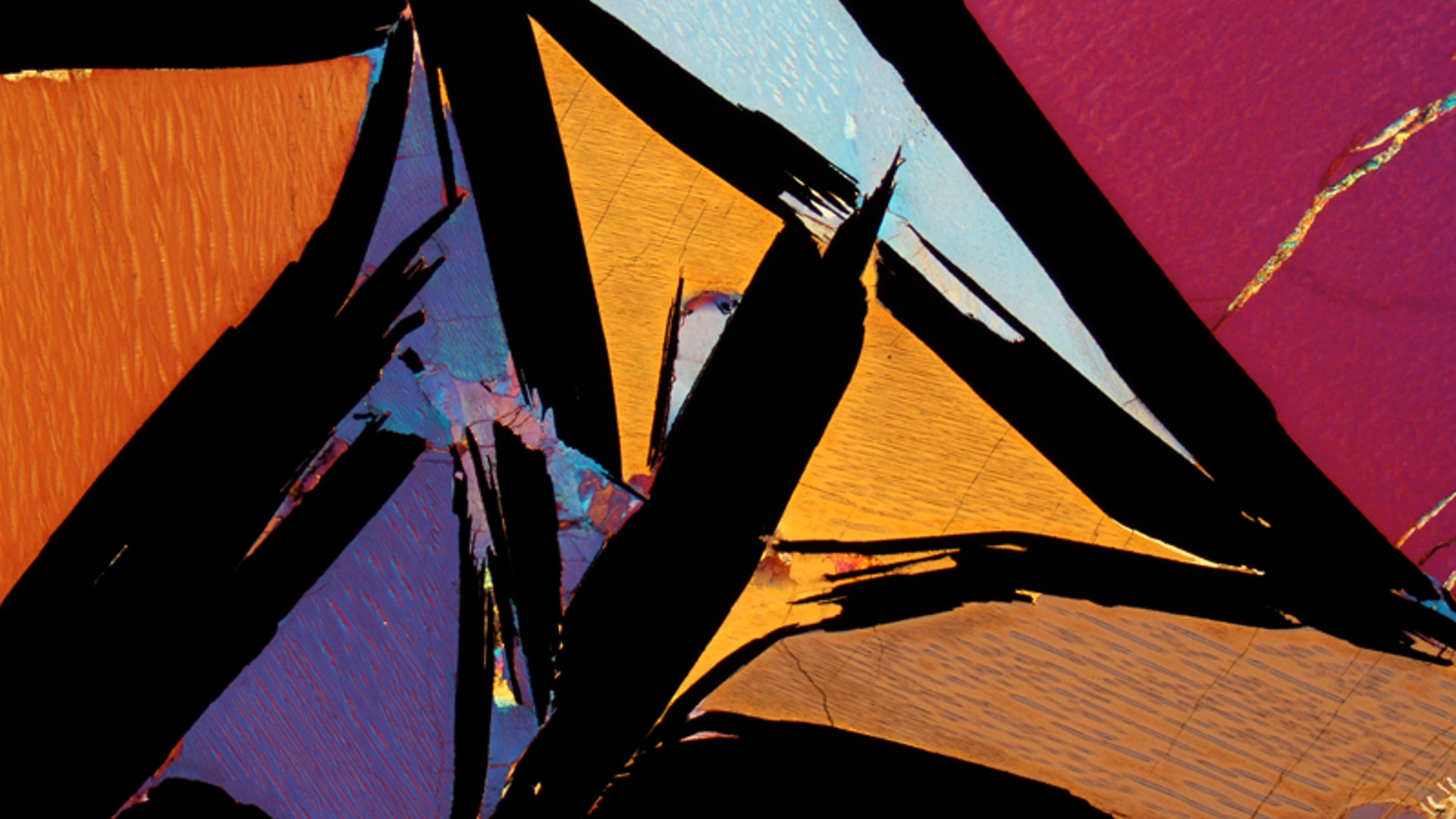
7th Place -- Gabriel Luna Nerve fibers from the retina of a mouse Technique: Laser confocal scanning, magnified 40X![8 Bernardo Cesare]() 8th Place -- Dr. Bernardo Cesare Coarse-grained rocks bearing graphite Technique: Polarized light, magnified 2.5X
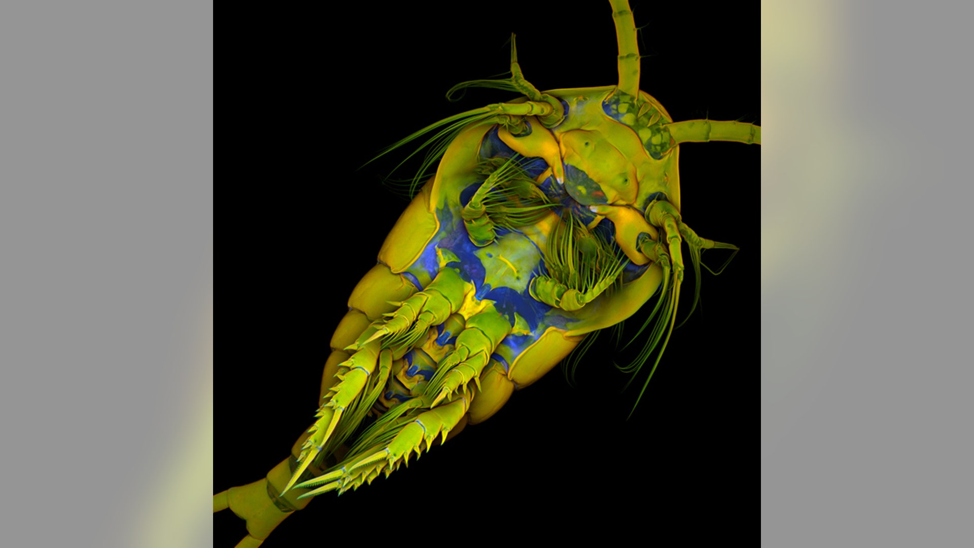
8th Place -- Dr. Bernardo Cesare Coarse-grained rocks bearing graphite Technique: Polarized light, magnified 2.5X![9 Jan Michels]() 9th Place -- Dr. Jan Michels The underbelly of a marine copepod Technique: Confocal, autofluorescence and congo red fluorescence, magnified 10X
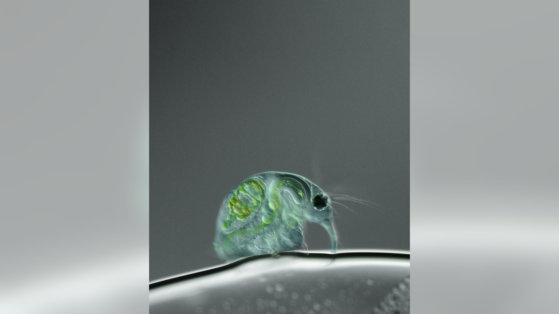
9th Place -- Dr. Jan Michels The underbelly of a marine copepod Technique: Confocal, autofluorescence and congo red fluorescence, magnified 10X![10 Joan Rohl]() 10th Place -- Joan Rohl Freshwater water flea Technique: Differential interference contrast, magnified 100X
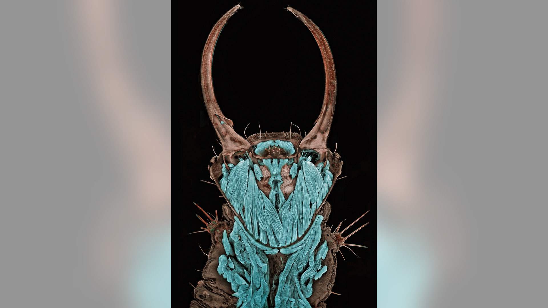
10th Place -- Joan Rohl Freshwater water flea Technique: Differential interference contrast, magnified 100X![11 Jan Michels]() 11th Place -- Dr. Jan Michels Front view of an ant head Technique: Confocal, autofluorescence, magnified 10X
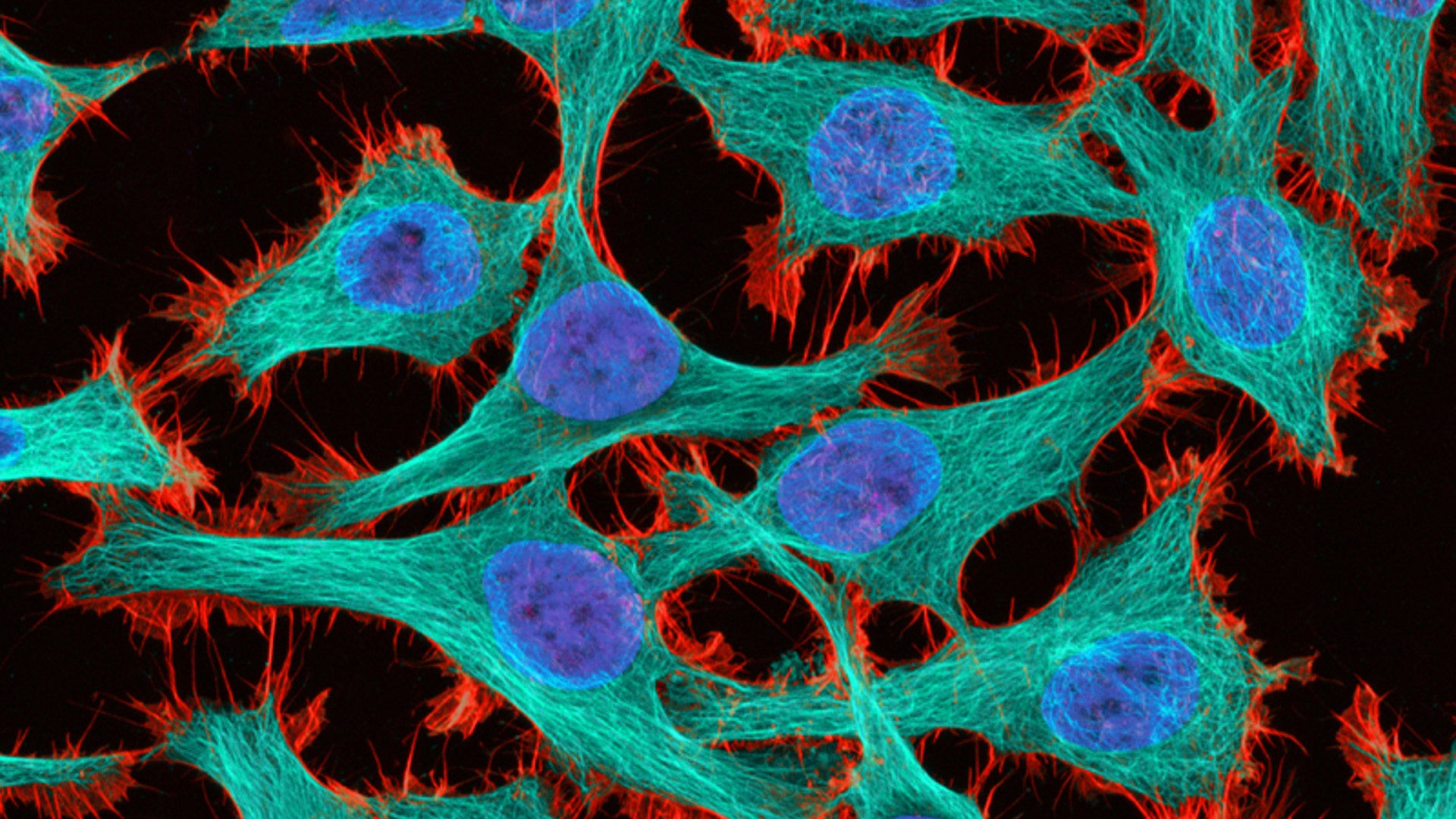
11th Place -- Dr. Jan Michels Front view of an ant head Technique: Confocal, autofluorescence, magnified 10X![12 Thomas Deerinck]() 12th Place -- Thomas Deerinck "Immortal" cancer (HeLa) cells Technique: 2-Photon fluorescence, magnified 300X
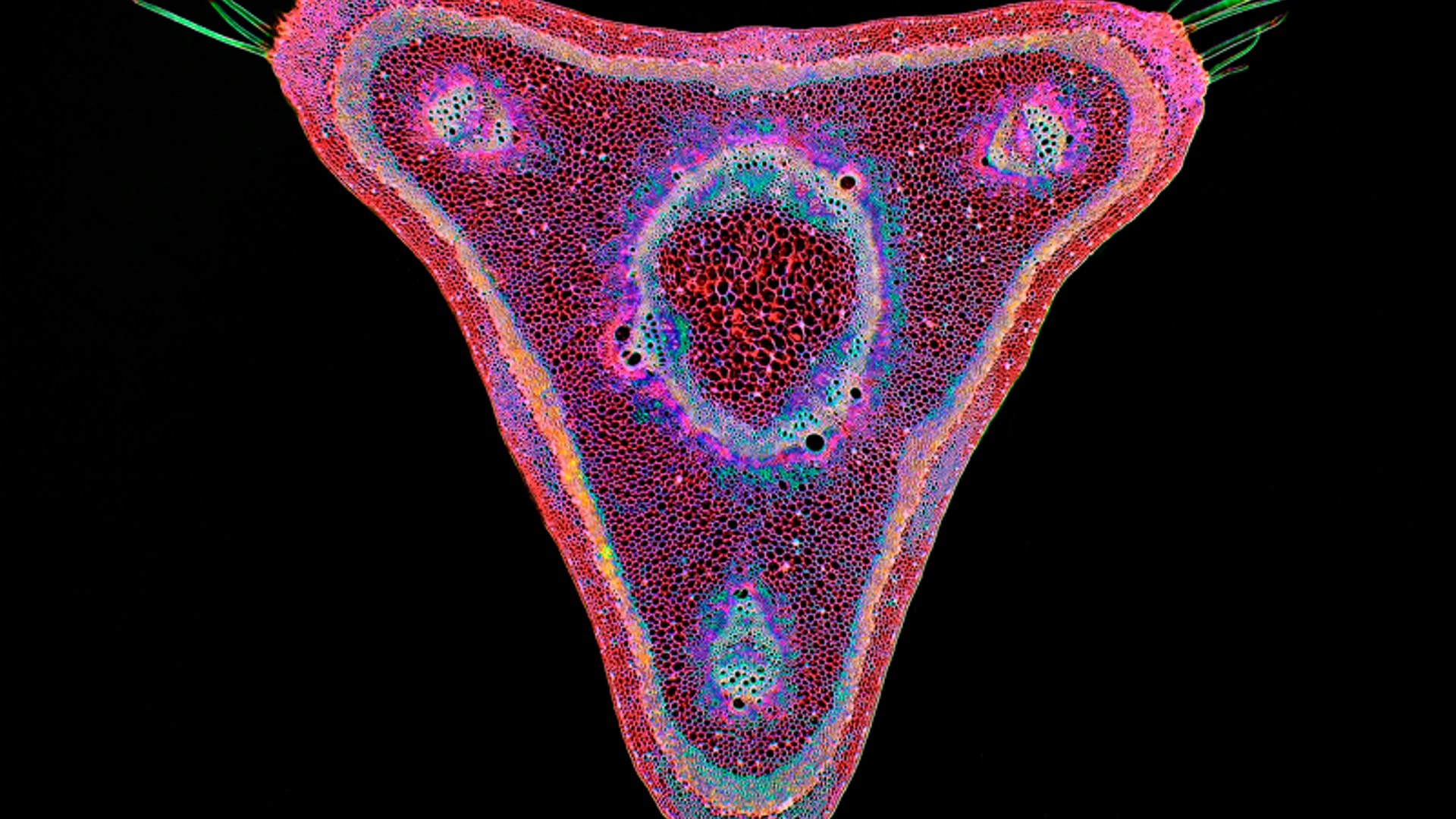
12th Place -- Thomas Deerinck "Immortal" cancer (HeLa) cells Technique: 2-Photon fluorescence, magnified 300X![13 Stephen S Nagy]() 13th Place -- Dr. Stephen S. Nagy A cross-section of a curare vine Technique: Brightfield, digitally inverted, magnified 45X
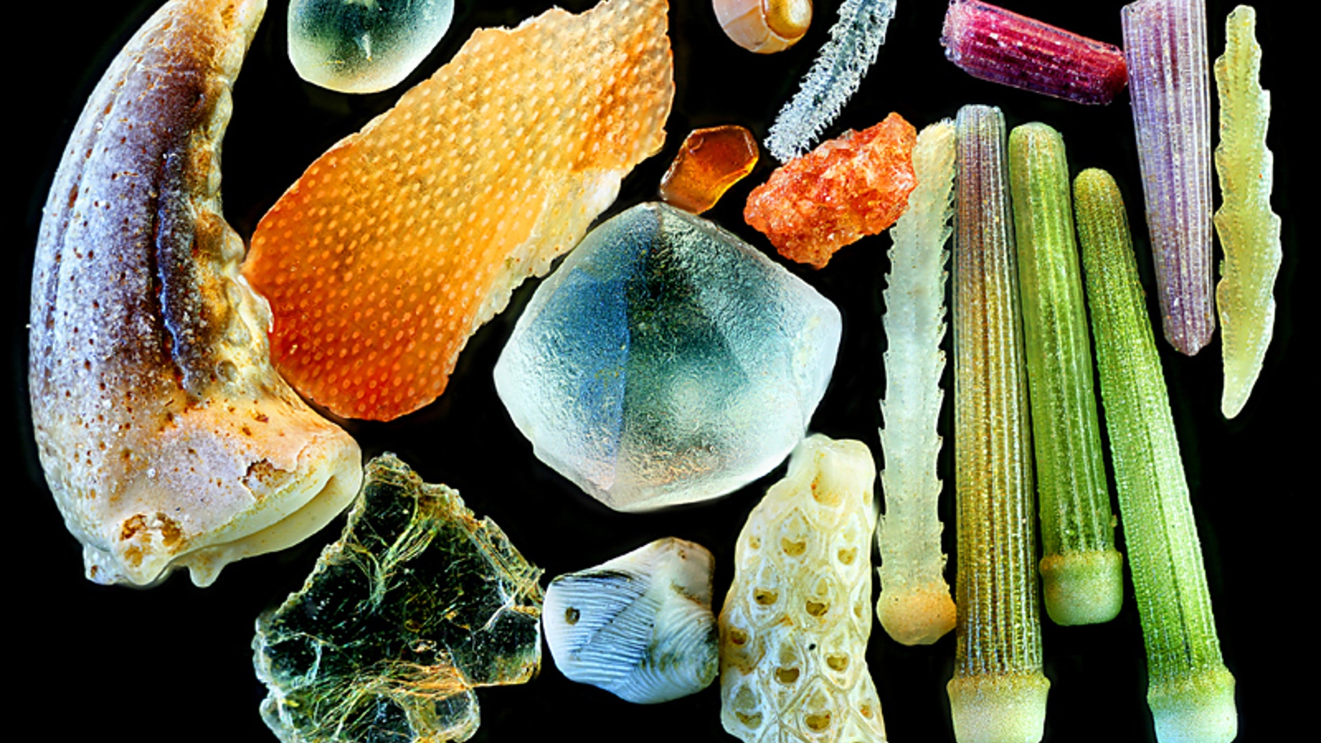
13th Place -- Dr. Stephen S. Nagy A cross-section of a curare vine Technique: Brightfield, digitally inverted, magnified 45X![14 Yanping Wang]() 14th Place -- Yanping Wang Various grains of sand Technique: Reflected light, magnified 4X
14th Place -- Yanping Wang Various grains of sand Technique: Reflected light, magnified 4X![15 James H Nicholson]() 15th -- James H. Nicholson A live specimen of lobe coral, its tissue pigmented with red fluorescence Technique: Epiflurescence with triple band (U/B/G) excitation, magnified 12X
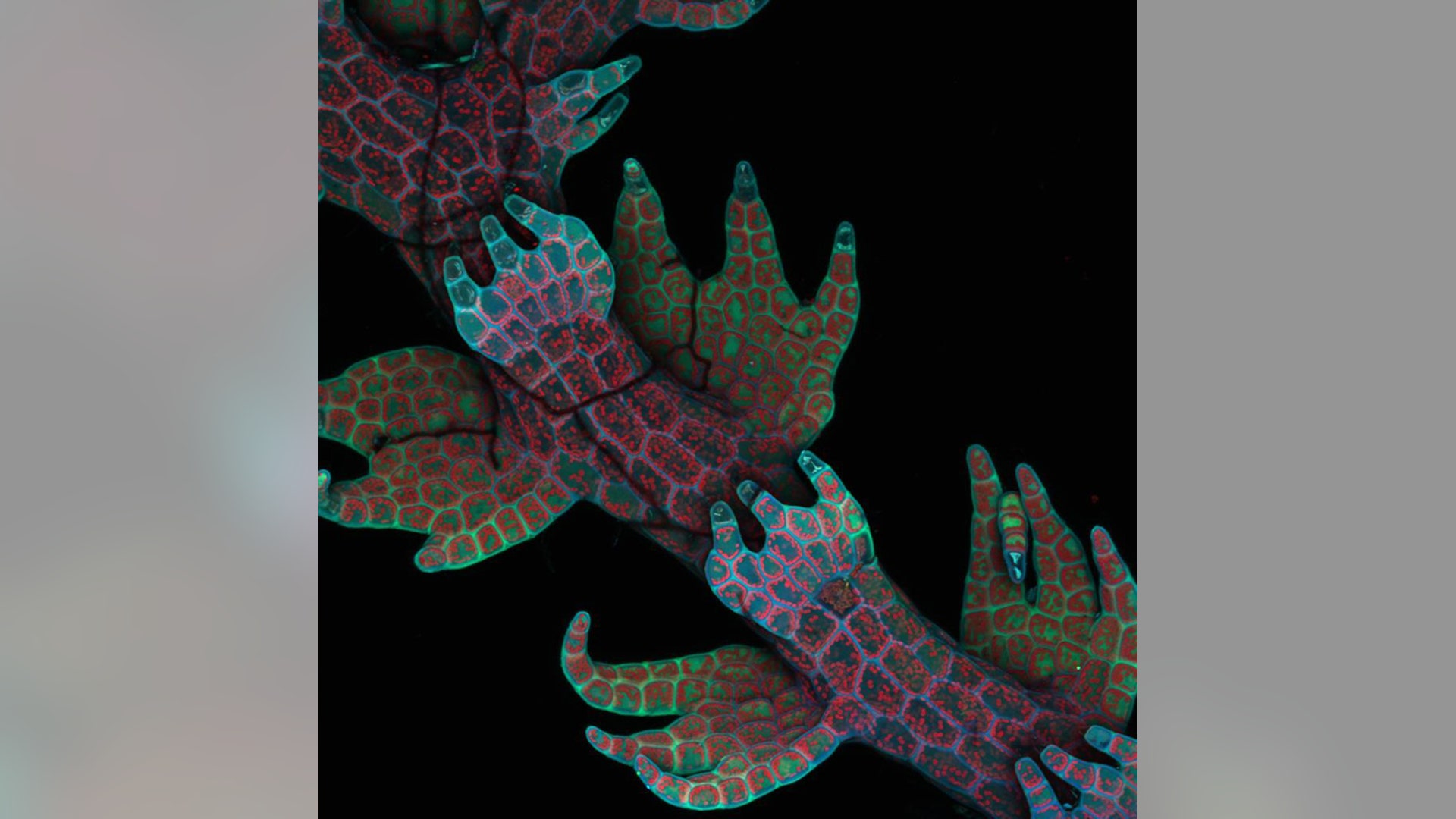
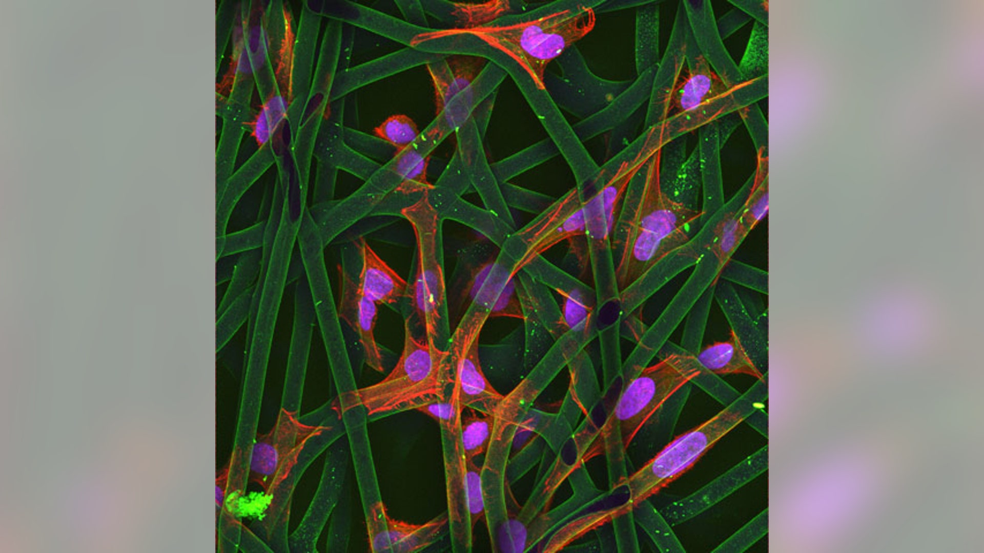
15th -- James H. Nicholson A live specimen of lobe coral, its tissue pigmented with red fluorescence Technique: Epiflurescence with triple band (U/B/G) excitation, magnified 12X![16 Christopher Guerin]() 16th Place -- Dr. Christopher Guerin Cultured cells Technique: Confocal, magnified 63X
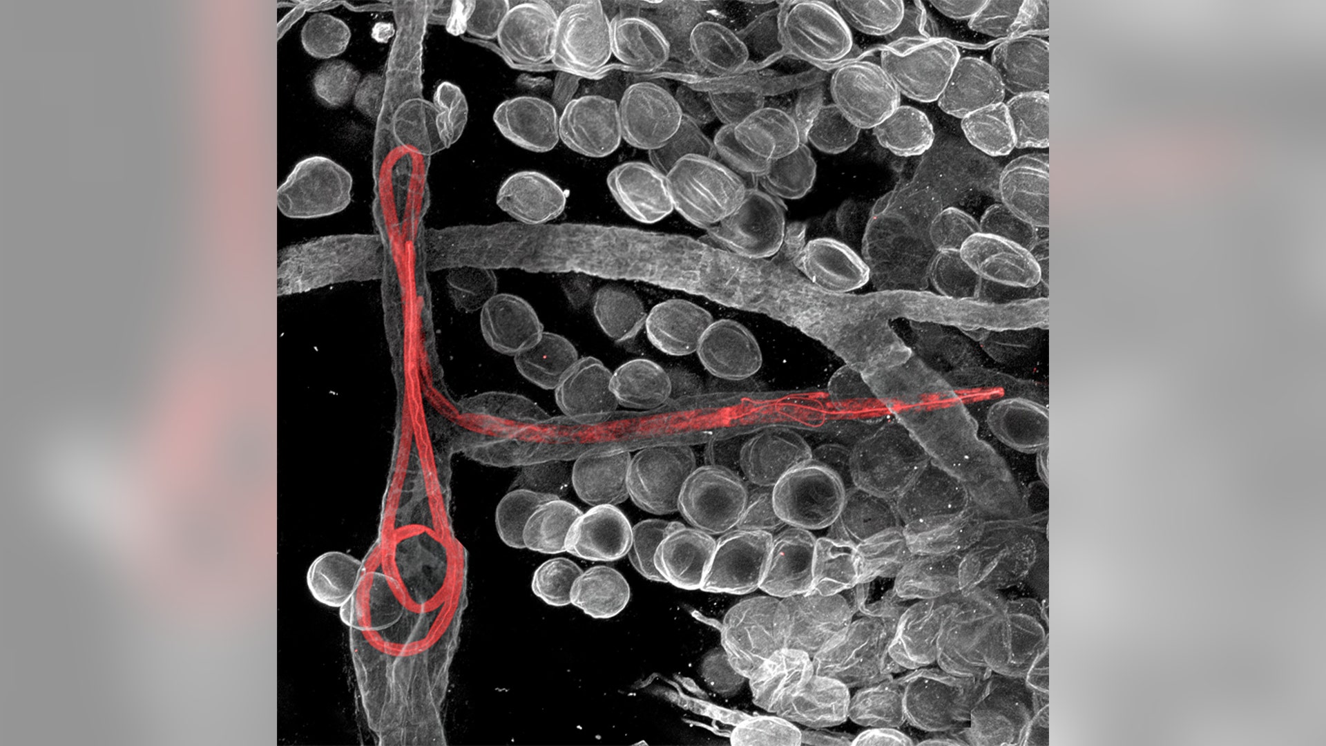
16th Place -- Dr. Christopher Guerin Cultured cells Technique: Confocal, magnified 63X![17 Witold Kilarski]() 17th Place -- Dr. Witold Kilarski Filaria worms inside the ear of a mouse Technique: Fluorescent confocal microscopy, magnified 150X
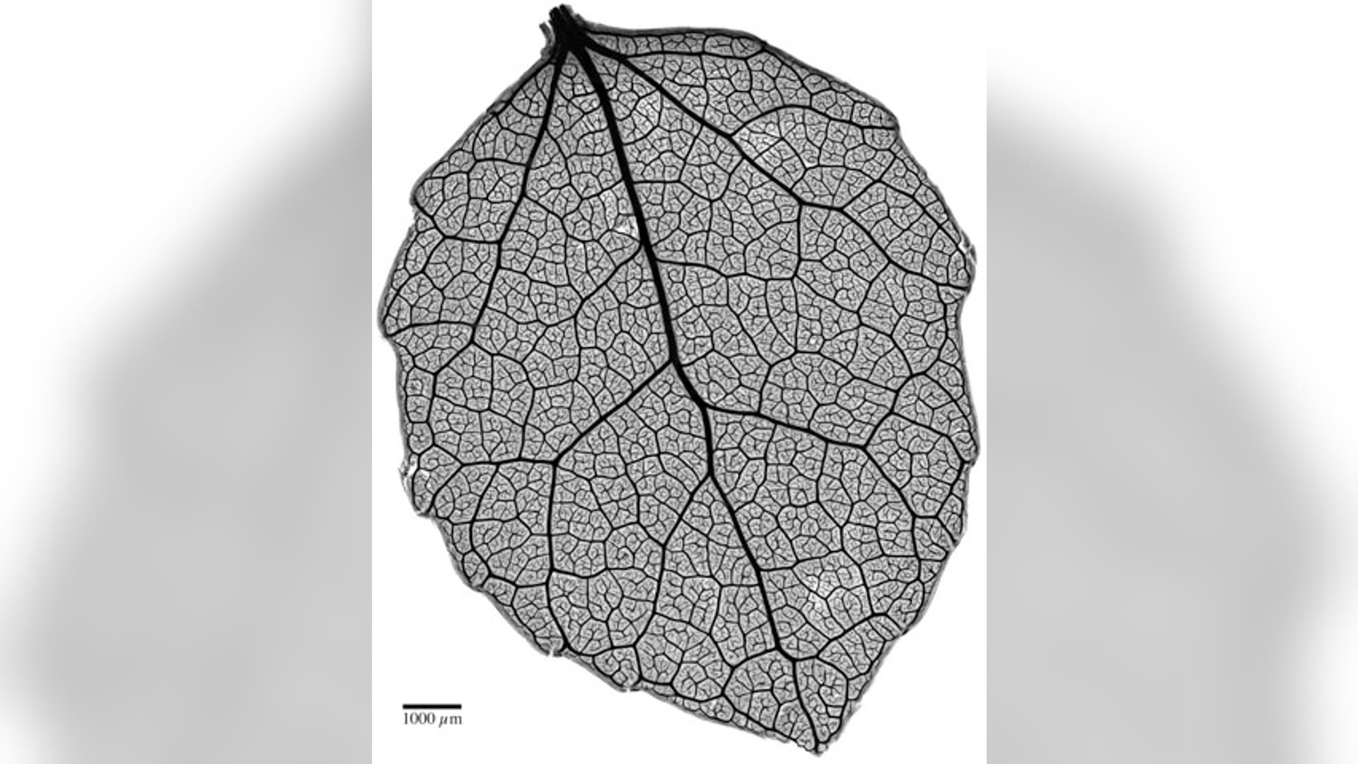
17th Place -- Dr. Witold Kilarski Filaria worms inside the ear of a mouse Technique: Fluorescent confocal microscopy, magnified 150X![18 Benjamin Blonder and David Elliot]() 18th Place -- Banjamin Blonder, David Elliot The venation network of a young quaking aspen leaf Technique: Brightfield image of safranin-stained tissue, magnified 4X
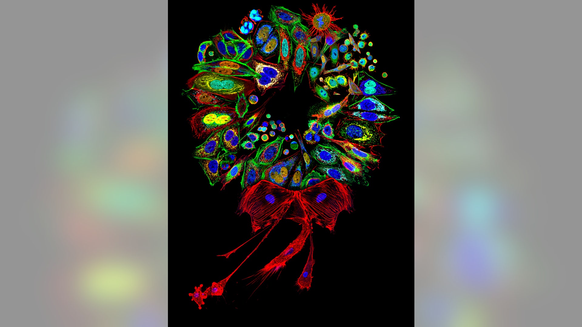
18th Place -- Banjamin Blonder, David Elliot The venation network of a young quaking aspen leaf Technique: Brightfield image of safranin-stained tissue, magnified 4X![19 Donna Stolz]() 19th Place -- Dr. Donna Stolz A collage of mammalian cells stained for various proteins and organelles, assembled into the shape of a wreath Technique: Single slice confocal cell mosaic, magnified 200-2000X
19th Place -- Dr. Donna Stolz A collage of mammalian cells stained for various proteins and organelles, assembled into the shape of a wreath Technique: Single slice confocal cell mosaic, magnified 200-2000X![20 Douglas Moore]() 20th Place -- Douglas Moore Dinosaur bone cells, unpolished and over 150 million years old Technique: Stereomicroscopy, fiber optics, magnified 42X
20th Place -- Douglas Moore Dinosaur bone cells, unpolished and over 150 million years old Technique: Stereomicroscopy, fiber optics, magnified 42XBest Microscope Photos From 2011 Nikon Small World Contest
The world as you've never seen it before: Nikon's annual Small World Competition showcases the year's best photography through the eye of a microscope. Here are the top 20 of 2011.


Move Forward
- Best Microscope Photos From 2011 Nikon Small World Contest


