Move Back
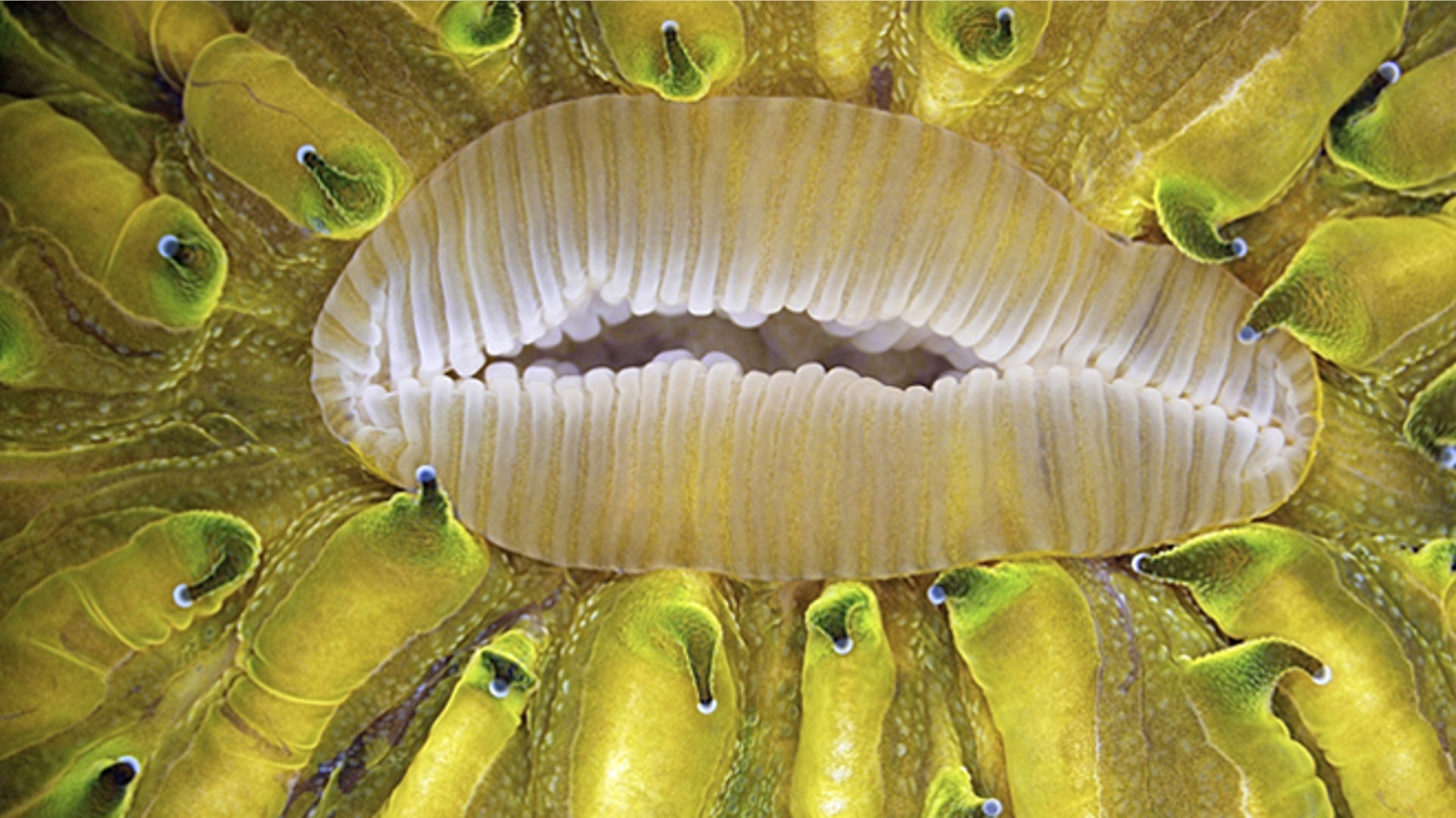
![2012_Olympus_Bioscape]() Sixth Prize: Live mushroom coral Fungia sp.Close-up of mouth during expansion. Captured using tungsten illumination; the green color is bright autofluorescence.
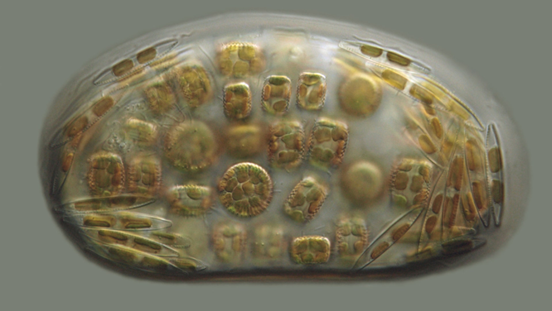
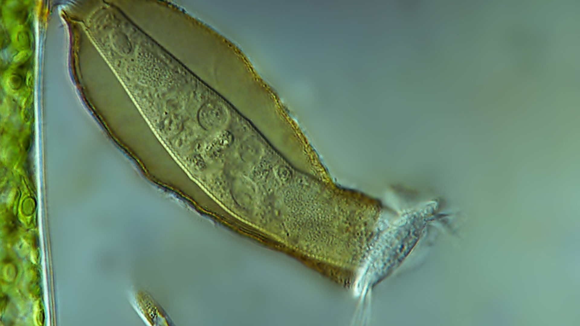
Sixth Prize: Live mushroom coral Fungia sp.Close-up of mouth during expansion. Captured using tungsten illumination; the green color is bright autofluorescence.![freshwater_diatom_cells]() Honorable Mention: Living freshwater diatom cells in a drop of water. Two species, are visible: Cyclotella meneghiniana (tablet shaped) and Nitzschia palea (long). Differential interference contrast. Victor Chepurnov, Algae R&D Office, Ghent, Belgium.
Honorable Mention: Living freshwater diatom cells in a drop of water. Two species, are visible: Cyclotella meneghiniana (tablet shaped) and Nitzschia palea (long). Differential interference contrast. Victor Chepurnov, Algae R&D Office, Ghent, Belgium.![freshwater_green_algae]() Honorable Mention: Various species of Desmids (freshwater green algae). Living specimens arranged as they naturally presented themselves on the slide. Image stack captured using polarized light at 100x. Marek Mis, Suwalki, Poland.
Honorable Mention: Various species of Desmids (freshwater green algae). Living specimens arranged as they naturally presented themselves on the slide. Image stack captured using polarized light at 100x. Marek Mis, Suwalki, Poland.![birth_of_bugs]() Honorable Mention: "Birth of bugs." Brightfield image stack. Frederic Labaune, Auxonne, France.
Honorable Mention: "Birth of bugs." Brightfield image stack. Frederic Labaune, Auxonne, France.![tunicate_larva]() Honorable Mention: Free-swimming tunicate larva. Darkfield illumination, magnification: 60x. Wim van Egmond, Rotterdam, The Netherlands.
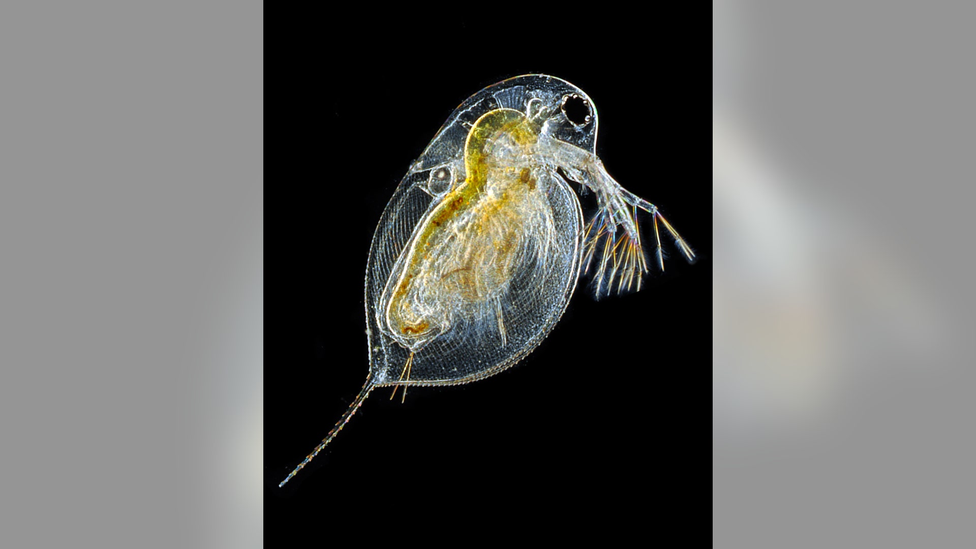
Honorable Mention: Free-swimming tunicate larva. Darkfield illumination, magnification: 60x. Wim van Egmond, Rotterdam, The Netherlands.![water_flea]() Honorable Mention: Daphnia (water flea), captured using image stacking. Michael Crutchley, Pembrokeshire, Wales, UK. Darkfield imaging.
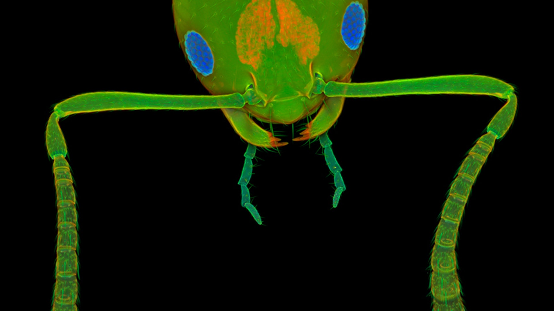
Honorable Mention: Daphnia (water flea), captured using image stacking. Michael Crutchley, Pembrokeshire, Wales, UK. Darkfield imaging.![worker_ant_frontal]() Honorable Mention: Worker ant. Frontal view of the head of a pharaoh ant (Monomorium pharaonis), a common indoor pest. Confocal microscopy. Jan Michels, Institute of Zoology, Christian-Albrechts-University of Kiel, Germany.
Honorable Mention: Worker ant. Frontal view of the head of a pharaoh ant (Monomorium pharaonis), a common indoor pest. Confocal microscopy. Jan Michels, Institute of Zoology, Christian-Albrechts-University of Kiel, Germany.![aspergillus_on_bread]() Honorable Mention: Aspergillus niger on bread, magnification 10x. Brightfield illumination. Chao Zhang, Beijing Language and Culture University, Beijing, China.
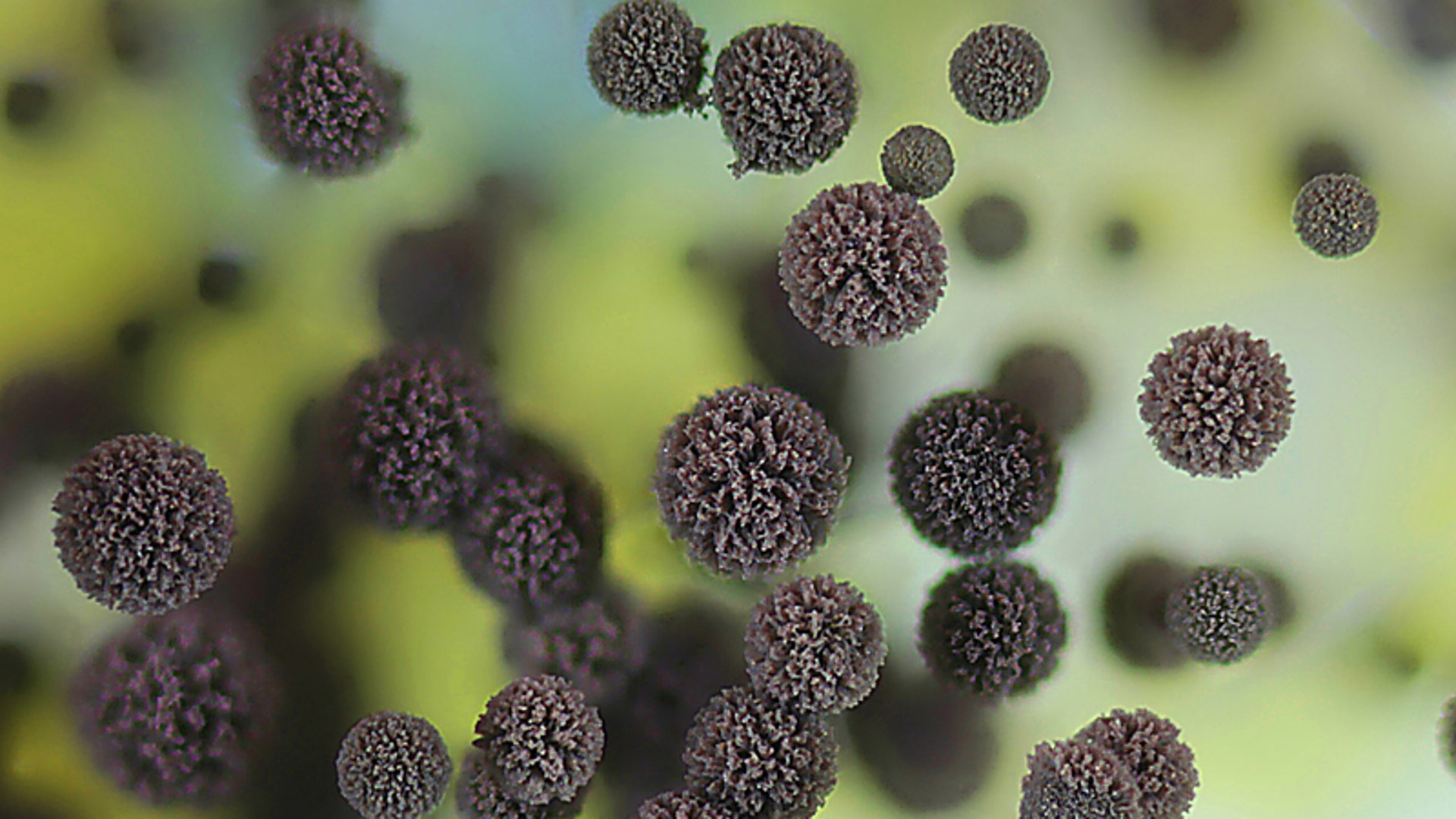
Honorable Mention: Aspergillus niger on bread, magnification 10x. Brightfield illumination. Chao Zhang, Beijing Language and Culture University, Beijing, China.![us_fern_spore]() Third Prize: A common East-coast US fern, Polypodium virginianum, showing a cluster of spore-filled sporangia and specialized protective hairs called paraphyses. Confocal microscopy. Igor Siwanowicz, HHMI Janelia Farm Research Campus, Ashburn, Virginia, USA.
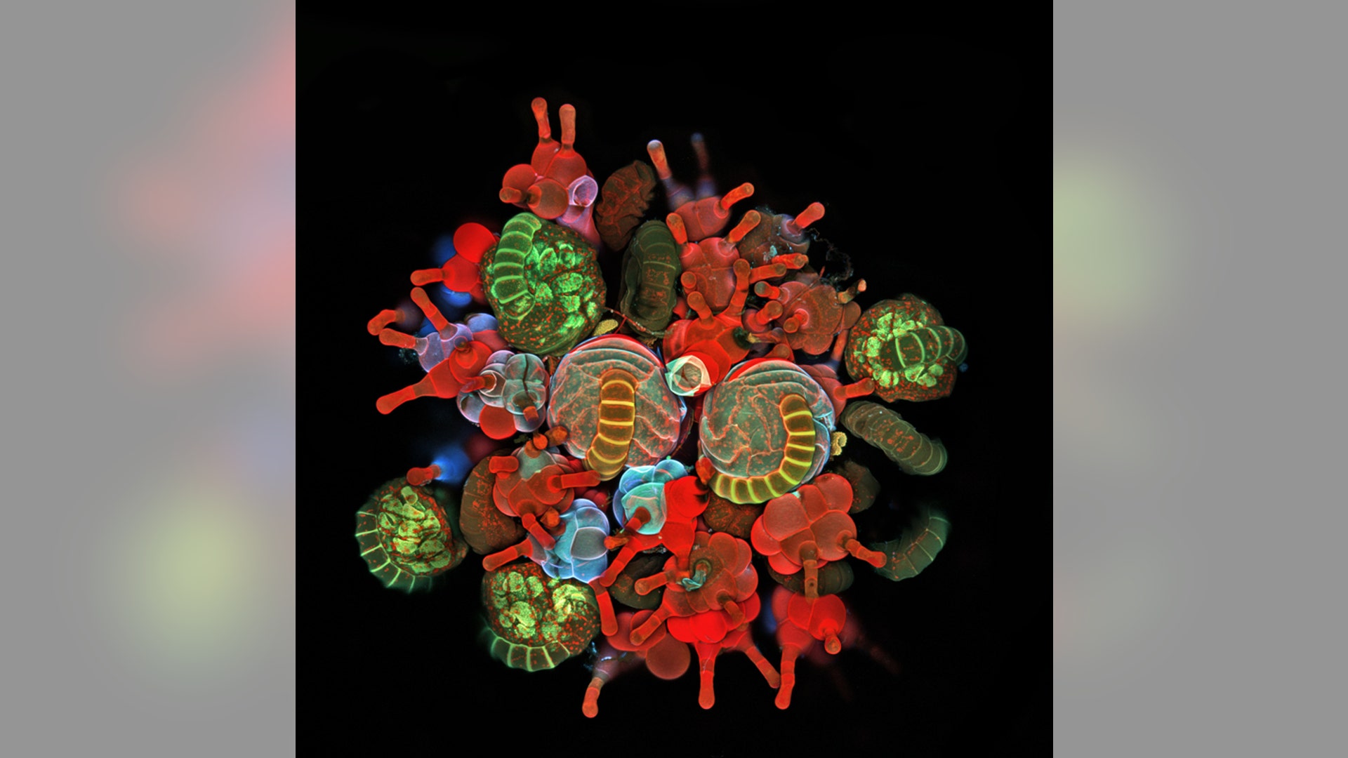
Third Prize: A common East-coast US fern, Polypodium virginianum, showing a cluster of spore-filled sporangia and specialized protective hairs called paraphyses. Confocal microscopy. Igor Siwanowicz, HHMI Janelia Farm Research Campus, Ashburn, Virginia, USA.![black_bread_mold]() Honorable Mention: Black bread mold (Rhizopus stolonifer) sporangia (300 micrometer). Image of the specimen in a Petri dish was captured using stereo microscopy. Csaba Pintér, Keszthely, Hungary.
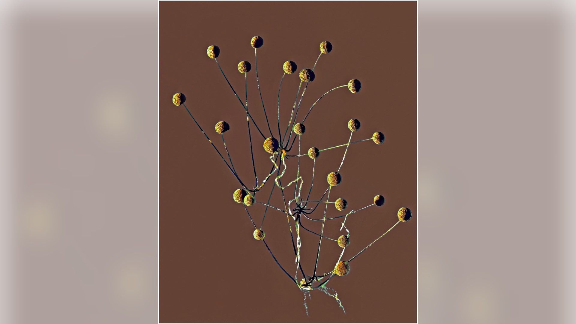
Honorable Mention: Black bread mold (Rhizopus stolonifer) sporangia (300 micrometer). Image of the specimen in a Petri dish was captured using stereo microscopy. Csaba Pintér, Keszthely, Hungary.![living_green_algae]() Honorable Mention: Living green alga Euglena mutabilis. Differential interference contrast, captured at around 900x. Gerd Guenther, Duesseldorf, Germany.
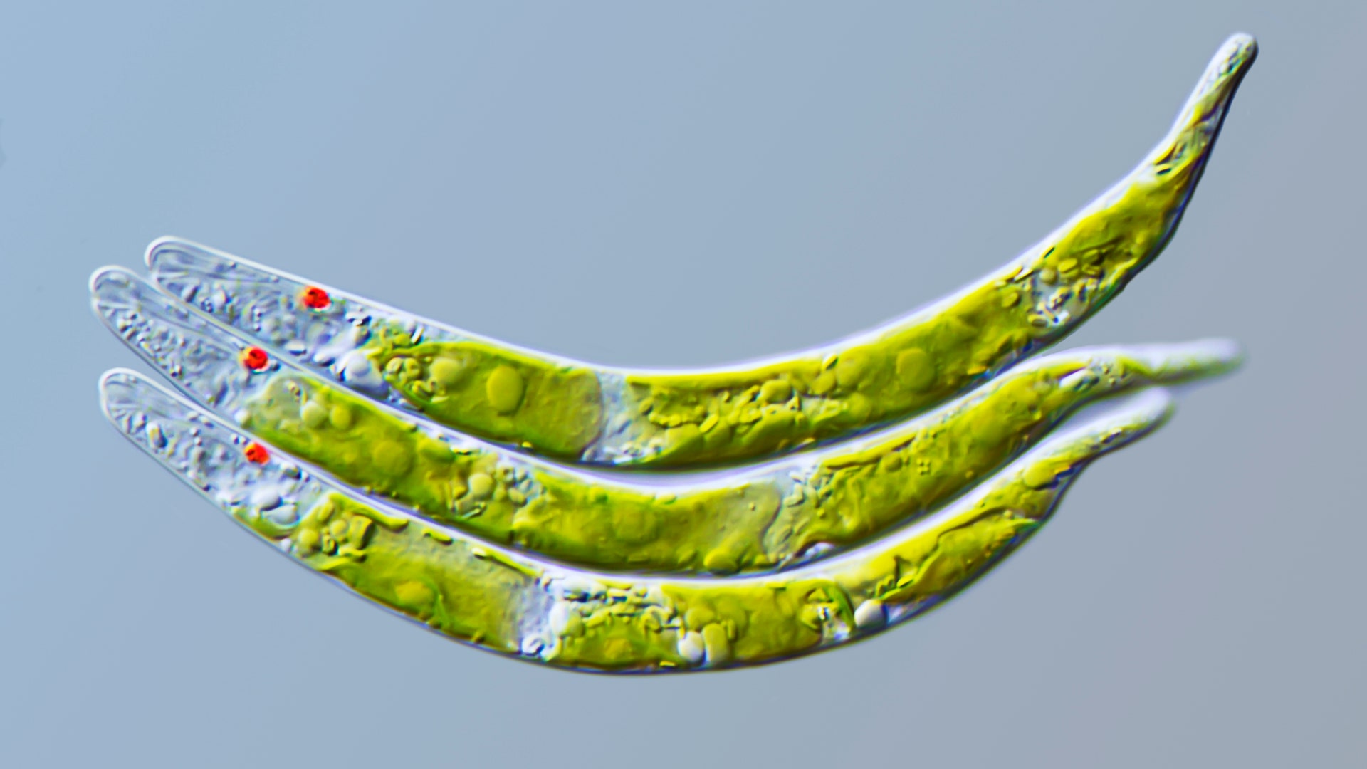
Honorable Mention: Living green alga Euglena mutabilis. Differential interference contrast, captured at around 900x. Gerd Guenther, Duesseldorf, Germany.![soldier_fly]() Honorable Mention: Soldier Fly (Stratiomyidae). Epi-illumination. Laurie Knight, Maidstone, Kent, UK.
Honorable Mention: Soldier Fly (Stratiomyidae). Epi-illumination. Laurie Knight, Maidstone, Kent, UK.![protozoan_pyxicola]() Honorable Mention: Protozoan Pyxicola captured using differential interference contrast at 1250x. Jacek Myslowski, Wloclawek, Poland.
Honorable Mention: Protozoan Pyxicola captured using differential interference contrast at 1250x. Jacek Myslowski, Wloclawek, Poland.![rotifer_stephanoceras]() Honorable Mention: Rotifer Stephanoceras. Darkfield illumination. Wim van Egmond, Rotterdam, The Netherlands.
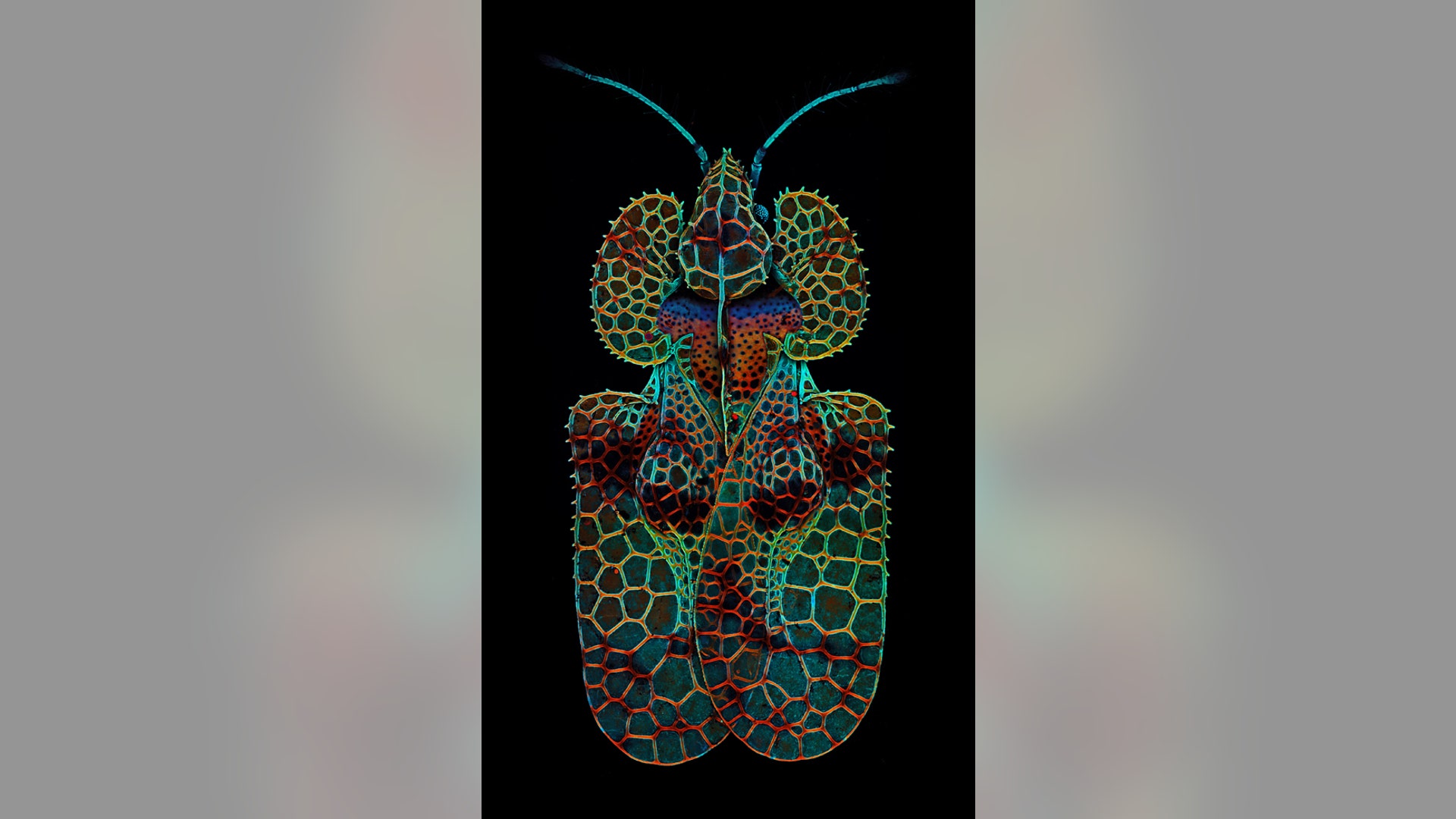
Honorable Mention: Rotifer Stephanoceras. Darkfield illumination. Wim van Egmond, Rotterdam, The Netherlands.![oak_lace_bug]() Honorable Mention: Oak lace bug (Corythucha arcuata), a common oak pest. Dorsal view of bug ca. 3mm long. Colors represent different structural properties of chitin and the varied affinity of the insect's exoskeleton to chitin-binding dyes. Eight stitched confocal images. Igor Siwanowicz, HHMI Janelia Farm Research Campus, Ashburn, Virginia, USA.
Honorable Mention: Oak lace bug (Corythucha arcuata), a common oak pest. Dorsal view of bug ca. 3mm long. Colors represent different structural properties of chitin and the varied affinity of the insect's exoskeleton to chitin-binding dyes. Eight stitched confocal images. Igor Siwanowicz, HHMI Janelia Farm Research Campus, Ashburn, Virginia, USA.Imperceptible organisms brought to life with microscope magic
The annual Olympus BioScapes Digital Imaging Competition recognizes the best microscope pictures around the world bringing to life entire worlds invisible to the naked eye.


Move Forward
- Imperceptible organisms brought to life with microscope magic


