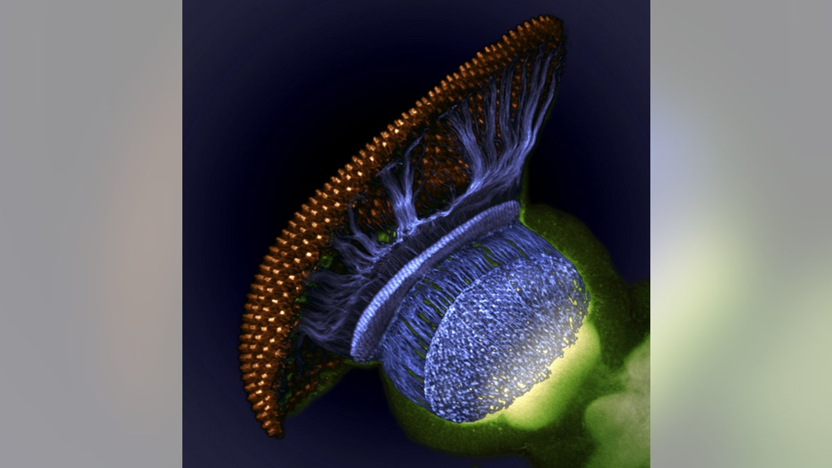
This space-y photo of a fruit fly (Drosophila melanogaster) visual system halfway through pupal development took home fourth prize. Image shows the fruit fly's retina (gold), photoreceptor axons (blue) and brain (green). (Dr. W. Ryan Williamson.)
A teensy bat embryo covering its eyes with wings, an ant carrying her larva and wobbly-looking newborn spiderlings are just a few of the winners of a photography contest honoring the little things in life — including babies, it seems.
The winners of the 2012 Nikon International Small World photography contest were announced today (Oct. 23), with a brightly colored image of the blood-brain barrier in a live zebrafish embryo awarded 1st place. The second-place winner in the small-world contest was a creepy-cute photograph of newborn lynx spiderlings by Walter Piorkowski.
Third place went to a wispy color-coded photo showing components of human bone cancer taken by Dylan Burnette of the National Institutes of Health in Bethesda, Md.
Nikon's Small World Competition, launched in 1974, honors those spectacular pictures taken through a microscope, called photomicrographs. Each year a panel of judges picks the top 20, plus honorable mentions. [See Winning Small World Photos]
"We are proud that this competition is able to demonstrate the true power of scientific imaging and its relevance to both the scientific communities as well as the general public," Eric Flem, communications manager of Nikon Instruments, said in a statement.
- Beauty Beneath the Microscope’s Lens
- Nikon’s Small World Contest 2012: Life as you’ve never seen it
- Fly Eyes to Stunning Snowflakes: the 2011 Nikon Small World Photo Contest
- Intricate salt grain wins ‘science as art’ competition
- Best Microscope Photos From 2011 Nikon Small World Contest
- Ordinary Is Extraordinary in 2011 Nikon Small World Photo Contest
[pullquote]
Some of the photo winners represent firsts, including the winning image taken by Drs. Jennifer Peters and Michael Taylor of St. Jude Children's Research Hospital. The duo developed a live transgenic zebrafish embryo that showed the development, in full color, of the blood-brain barrier — a structure that keeps material in the bloodstream from entering the brain. The image is thought to be the first showing the formation of the blood-brain barrier in a live animal.
And then there were the unrecognizable. An image that resembles more a mushroom-shaped spacecraft lifting off than a humble fruit fly (Drosophila melanogaster) took home fourth place. The space-y image, by W. Ryan Williamson of the Howard Hughes Medical Institute, reveals the fly's visual system halfway through pupal development. Another surreal image, this one of the fruit fly's eye organ, landed seventh place. The image was taken by the University's of Utah's Michael John Bridge.
A more grounded image took fifth place: University of Valencia, Spain, geologist Honorio Cócera-La Parra captured a close-up of the mineral cacoxenite, made up of iron aluminium phosphate, from La Paloma Mine in Spain.
The winning photomicrographs will be displayed in a color calendar and in a national museum tour. And while the judges have chosen this year's winners, Nikon is also hosting a popular vote on Facebook that runs through Nov. 13 and lets the public to choose their own winners.
Copyright 2012 LiveScience, a TechMediaNetwork company. All rights reserved. This material may not be published, broadcast, rewritten or redistributed.

Get a daily look at what’s developing in science and technology throughout the world.