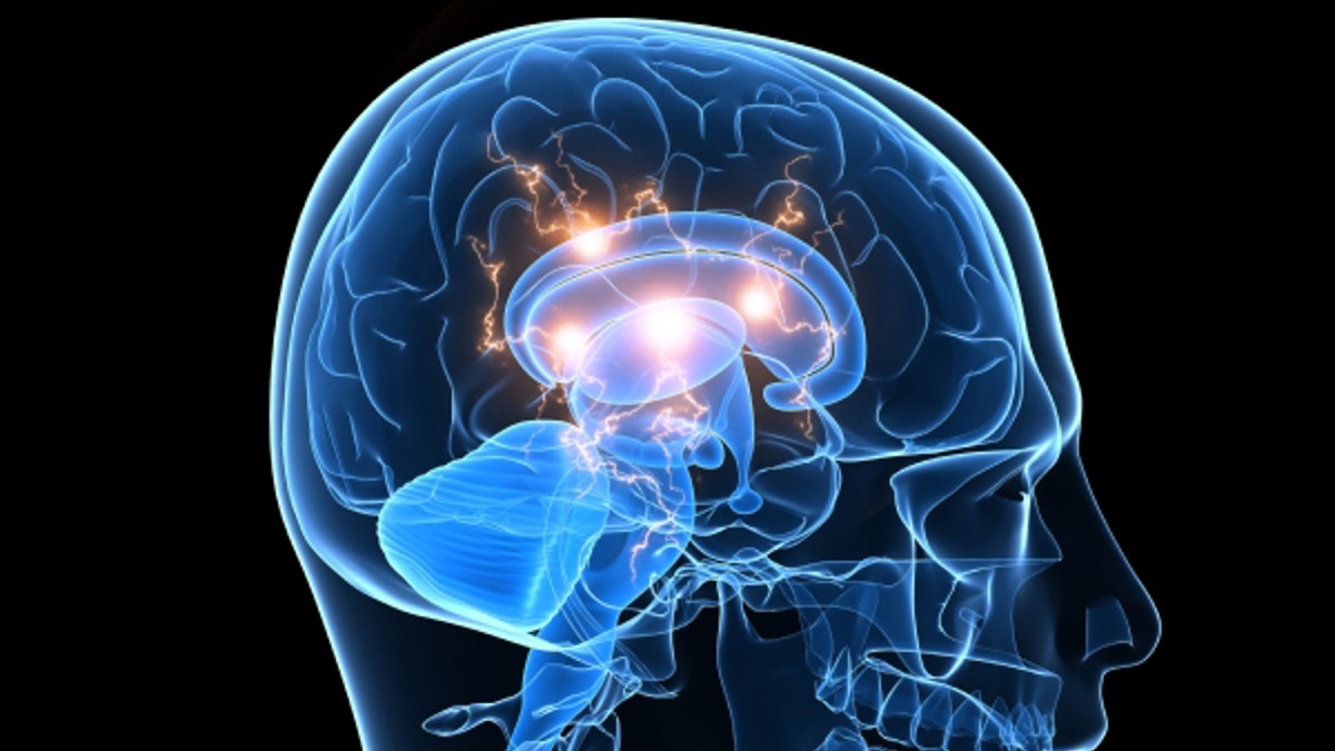
For decades, conducting an autopsy was the only way for doctors to determine if an Alzheimer’s patient had an accumulation of beta-amyloid plaques in the brain – a major hallmark of cognitive decline.
But over the past few years, brain imaging using an experimental radioactive dye has helped physicians confirm the presence of these plaques while patients are still alive. Now, a new multicenter study has confirmed that this type of scanning can detect early evidence of Alzheimer’s disease, predicting future impairment among patients with little to no symptoms.
The radioactive dye, florbetapir (AMYViD), works like a chemical stain in the brain. Once injected into the body, florbetapir binds and sticks to the brain’s beta-amyloid plaques, helping to estimate the extent of plaque buildup throughout the brain’s regions. Then, through positron emission tomography (PET) scanning, a radioactive tracer looks for chemicals in the dye and produces an image highlighting the positioning of the plaques in the brain.
“[It] helps doctors when they encounter someone with progressive memory problems, and they’re trying to determine the cause,” lead author Dr. P. Murali Doraiswamy, professor of psychiatry and director of the neurocognitive disorders program at Duke University, told FoxNews.com. “…If this PET scan comes back positive, it would support an [Alzheimer’s] diagnosis. If the PET scan comes back as negative, the doctor needs to look at other causes. The scan by itself is not diagnostic of any condition, but it can help doctors increase the accuracy of a diagnosis.”
Florbetapir was approved by the U.S. Food and Drug Administration in 2012 as a way to estimate plaque density in individuals suffering from cognitive impairment; however, the dye has not yet been approved to predict the development of dementia, and it is not used as a screening tool in people with normal cognition.
To better understand florbetapir’s ability to forecast Alzheimer’s disease, Doraiswamy and other researchers from Duke University, Harvard Medical School and more scanned the brains of 152 adults over the age of 50. The participants were separated into three groups: 69 had normal cognition, 52 had mild memory problems, and 31 had been clinically diagnosed with Alzheimer’s disease.
The researchers periodically conducted PET scans on the study participants for a total of three years, while also giving them a series of cognitive exams to assess their intellectual abilities. The PET scans were rated as positive for a significant amount of plaques or negative for very little to no plaques.
“Our goal was to see if a baseline PET scan would predict who would decline faster and who wouldn’t,” Doraiswamy said. “…And people with positive scans declined cognitively at a much faster rate…People with mild memory problems with a positive scan had a three times greater rate of progressing to Alzheimer’s [than people with negative scans].”
Of the participants, 35 percent with plaque-positive scans who started the study with mild cognitive impairment progressed to Alzheimer’s, compared to only 10 percent of participants with no plaques. Conversely, the negative scans also held a predictive value: 90 percent of participants with mild memory problems but no plaques did not progress to Alzheimer’s disease.
According to Doraiswamy, more evidence is still needed to confirm florbetapir as a predictive tool, but he said their results are encouraging – especially given the fact that the dye is a mostly benign chemical.
“It’s a radioactive dye that uses an extremely low amount of radioactivity and has an extremely low half-life,” Doraiswamy said. “…The PET scan actually has more radioactivity, and if someone is getting a single PET scan for the year, that’s acceptable exposure.”
Doraiswamy also noted that PET scanning with florbetapir could open up many opportunities for developing new drugs that clear plaques from the brain. With this test, physicians can identify individuals with plaques, enroll them in a trial of an experimental drug and then repeat PET scans to see if the drug is successfully clearing plaques.
Overall, Doraiswamy said this scan adds to an ever-growing arsenal of tools physicians can use to better understand Alzheimer’s disease – a condition that currently affects more than 5 million Americans and is the sixth leading cause of death in the United States.
“For diagnosis, there are MRIs, blood tests, memory tests, and now PET scans,” Doraiswamy said. “No one test can diagnose, but PET scans can tell if someone has plaques in the brain. Before this scan was available, when you saw a patient in the office, you didn’t really know if they had plaques. Now with this scan, you can tell them ‘I’m seeing a positive scan, so the odds are you will progress to Alzheimer’s.’”
The research was published online in Molecular Psychiatry.

Stay up-to-date on the biggest health and wellness news with our weekly recap.